Should wisdom teeth be preserved?
We think yes.
Although wisdom teeth are not involved in the process of chewing food, they can still benefit a person. There must be clear medical indications for their removal. And now we will provide you with another proof of the benefits of your own teeth-eights. Our patient Kim is about 20 years old, she is from Korea and lives in Moscow, as her parents work at the Korean embassy. I contacted the German Implantology Center due to inflammation of tooth 4.6 – the lower right six. The tooth was destroyed
, an abscess developed. There was a temporary filling on the tooth (placed in another clinic):
Where a temporary filling was placed, she was told to remove the tooth. That's why Kim came to us with one simple goal - to atraumatically remove the six, which began to greatly bother and hurt her. After doing a CT scan and looking at the tooth, we offered her an option, which, upon hearing, Kim at first did not believe her ears. Our proposal was to remove the six and immediately in its place... transplant its lower right wisdom tooth! This is called autotransplantation surgery.
When do baby teeth start falling out?
Baby teeth begin to form while the baby is still in the mother's womb. Moreover, their number is not 32, as in an adult, but only 20. Baby teeth usually fully erupt by the age of 2. At the age of approximately 6 years, the replacement of baby teeth with permanent teeth begins to occur. In this case, 20 existing teeth fall out, and the remaining 12 erupt. This usually happens in the following order:
- The deciduous incisors are usually the first to fall out. The roots of the teeth begin to thin out, and when the baby reaches a certain age, the incisor falls out. For central incisors it is 6 years, for lateral incisors - 7 years.
- Then the molars begin to fall out. These baby teeth are divided into first and second teeth. Loss of first molars occurs between 7 and 10 years of age. The second molars are the last to fall out, between the ages of 9 and 13.
- In the interval between the loss of the first and second molars, loosening and loss of the upper and lower canines occurs. This occurs between the ages of 9 and 11 years.
It should be remembered that the process of replacing baby teeth with molars is individual, and the indicated timing is not necessarily suitable for all children.
Wisdom tooth transplant as a way to replace a defect
Of course, Kim was very surprised, but we showed her photographs of successful operations (and we have all of them successful), apparently our persuasiveness and the patient’s desire played a positive role in making the decision to transplant the eighth tooth. She was happy and agreed to the operation.
Moreover, in this case we did not even have to make a stereolithographic model of the tooth, since all surgical procedures (extraction and transplantation) were performed on the very first day of her visit to our clinic.
Dental transplant surgeries typically require several days of preparation, as we describe in the following clinical case study. But in this case, taking into account the current clinical situation and relying on our many years of successful experience in tooth transplantation, we were able to transplant tooth 8 into the place of tooth 6 as quickly as possible. We had all the indications for current dental transplantation.
The donor was an impacted (completely hidden under the gum) wisdom tooth 48. In fact, as we joked, it was a neighbor tooth to the problematic six. That is, the tooth grew and grew for 20 years, waited in the wings, and that hour struck! Now this donor tooth is for another 30 years
at least he will live in a new place.
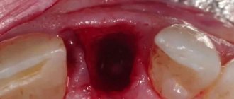
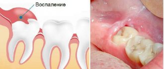
If you look at the sixth tooth from the side, you can see bluish, literally purple gums
:
This color of the gums is due to the developed inflammatory process. The patient developed destruction of bone tissue against the background of advanced periodontitis. There was a crack in the root of the sixth tooth.
Why does a person's teeth change?
The structure and development of the dentition in different animals differs significantly from each other. In vertebrates and some species of reptiles, for example, teeth change several times in their lives, and over time, a new one grows in place of the lost tooth; in rodents, permanent teeth immediately grow. This is due to the characteristics of the body, residence and nutrition of representatives of the animal world.
There are several species of mammals whose teeth change once in their lifetime during childhood. This is necessary not only for the growth of the body, but also for survival - after the baby stops feeding on mother’s milk, its life largely depends on the capabilities of the dental apparatus. Humans can also be classified as mammals in this category, so changing teeth is an integral process of growing up for them.
The difference between transplanting your own tooth and implantation with a dental implant
By the way, for your transplanted tooth, the presence of an inflammatory process in the soft and bone tissues does not matter, because all the tissues there are their own and, naturally, the body will protect its transplanted tissues. That is, the body does not treat the transplanted tooth as “friend or foe,” which always happens in the case of tooth implantation with a titanium or zirconium implant.
If there is an inflammatory process, an artificial implant cannot be placed in this place. And you can put your own tooth, because it has the so-called. the ligament of the tooth that has all the cells that will fight.
Naturally, after the tooth extraction, we processed everything as much as possible, carefully cleaned and rinsed it.
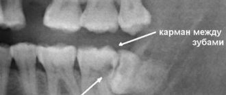
In this photo we have begun to treat the wound. It can be seen that there was an inflammatory process in the gums, the bone tissue was partially but severely destroyed. Signs of bone tissue destruction are clearly visible on computed tomography:
By the way, even if the bone tissue is destroyed, when we transplant our own teeth, we do not need bone grafting; there is no need to replant additional bone. And that's why. The tooth itself nourishes and regenerates. Even if there were no front wall of the tooth, if there were no vestibular buccal wall, if you transplant such a tooth, then it is restored from its ligament, even the bone grows.
Therefore, if there is a choice between transplanting your own tooth and implanting a dental (titanium, zirconium) implant, it is better to opt for transplanting your own tooth.
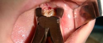
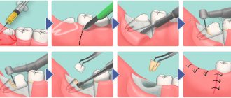
Let's begin atraumatic wisdom tooth removal
In 40 seconds
We made an incision in the area of the donor tooth, which we planned to remove and replant in place of the previously removed six. We carefully removed the wisdom tooth, placing it in a saline solution for a short time:
Many people wonder: do wisdom teeth have roots? Yes, of course there is – this is a molar. We draw your special attention to the fact that this wisdom tooth 48 has not yet finished growing its own roots, its maturation is not yet complete. The growth zone of the root of the lower wisdom tooth is preserved and this tooth subsequently, after 2 weeks, does not require nerve removal! That is, it not only grows in, but also completely becomes accustomed to its nerve.
a powerful ligamentous apparatus he has.
, which provides high survival potential. Indeed, this tooth has taken root, and besides, it has a living nerve.
Transplantation of a wisdom tooth into place 4.6
The donor tooth was clearly placed in the place of the problematic sixth tooth. It was as if he had been there all his life: ideal external dimensions, the wisdom tooth was transplanted to the place of the removed one without any external grinding. Ideal autotransplantation operation.
The implanted tooth is tightly sutured
to ensure maximum tissue contact with each other and a normal healing process.
Removal of baby teeth in children
It all depends on the number of lost baby teeth, their location in the jaw and how much time is left before the permanent tooth erupts. If it is three or four years, the more likely it is that the permanent tooth will be delayed or will not grow. The hard and compact bone tissue formed above the tooth germ will be difficult for him. The opposite is true when the time to a supposedly permanent dentition is quite short. Then the eruption of the permanent tooth usually occurs earlier than it would have happened naturally.
Premature loss of primary teeth can also interfere with alveolar bone growth. A child who does not have teeth cannot bite properly, and the forces generated during this activity are not transmitted to the bones and muscles, which does not contribute to their proper development.
Losing two baby teeth on one side, such as the fourth and fifth, can lead to other consequences. With such extensive missing teeth, the occlusal forces between teeth that shape and guide the growth of the alveolar bone are compromised. Thus, growth is uneven and is expressed in the vertical layering of bone where there are no teeth.
Premature loss of a molar, that is, the primary five, always leads to an internal (i.e. forward) displacement of the fixed six. This is a common problem that causes the dental arch to shrink. As a result of this phenomenon, there may not be enough space for other molars. Consequently, crowding of teeth occurs, this feature looks unsightly when there is no room for the canine tooth and it grows outside the dental arch or does not grow at all because due to lack of space, it gets stuck in the bone.
Premature loss of incisors and primary canines, that is, the upper front teeth, contributes to poor jaw bone development, which can lead to malocclusion. For example, if the jaw is underdeveloped compared to the lower jaw, an anterior transverse bite will form (the lower front teeth will overlap the upper ones).
Speech problems may occur in children with severely defective primary teeth, especially the front incisors. Such deficiencies do not contribute to the correct position of the tongue on the palate, so there is a possibility of interdental lisp. As a rule, after the removal of a baby tooth, a child needs 1-2 years of visiting a speech therapist or dentist.
Missing teeth and speech defects not only affect health, but also have a psychological aspect: the child may have low self-esteem, be secretive or shy because of his appearance. Children's baby teeth are truly worth caring for. Oral hygiene is equally important at every stage of life.
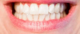
After autotransplantation 2 weeks
Patient Kim came to us after 2 weeks
for medical examination and suture removal:
Nothing bothered her anymore. The tooth had acceptable physiological mobility. The gums have become lighter. Of course, all the purple color is not gone yet, but she has already become more pink.
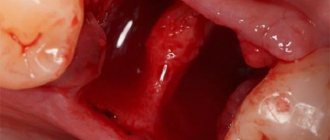
Pay attention to the white area - this is not pus, but fibrin plaque, which we will talk about in more detail below.
The picture shows that the donor tooth has clearly positioned itself in the position of the extracted tooth 46:
Our Kim has a new six molar. Both the patient and the medical staff of the Research Center are very pleased with the outcome of the tooth transplantation operation.
How are baby teeth different from molars?
A human tooth consists of three main parts: the visible part (crown), the root, which is recessed into the gum tissue, and the neck, located between them. Despite the fact that the general structure and functions of primary and permanent teeth are similar, there are still differences between them:
- the surface shade of temporary teeth is white, while permanent teeth are yellowish;
- the tissues of the molars are denser and harder, and the pulp is much larger;
- the shape of the milk teeth is flat and wide, the molars are quite long (the width is significantly less than the length);
- The roots of baby teeth are thinner and shorter than those of permanent teeth.
During the process of loss of temporary teeth and the appearance of permanent teeth, the child’s jaw actively grows, and by the time it ends it reaches an adult size - thanks to this, 32 permanent teeth grow in place of 20 temporary teeth.
About fibrin and fibrin plaque
Fibrin
- a high-molecular, non-globular protein formed from fibrinogen, synthesized in the liver, in the blood plasma under the action of the enzyme thrombin; has the form of smooth or cross-striated fibers, the clots of which form the basis of a blood clot during blood clotting. Thus, fibrin promotes rapid wound healing by forming fibrin plaque.
Fibrin plaque
- a natural phenomenon that occurs in the oral cavity in the area of surgical intervention (at the site where a tooth was removed, stitches were applied, etc.). It occurs on the 3-4th day after surgery in the oral cavity. And this is one of the normal and inevitable stages of healing of a hole covered with a blood clot. This is followed by tissue restructuring, covering the wound with protective epithelium and restoration of normal mucosa.
Patients often confuse fibrin plaque with pus, causing them to worry and call their dentists.
Why does fibrin plaque occur?
Healing of a wound in the oral cavity occurs in a moist environment. If you have a cut on your skin, once the bleeding stops, the wound will be covered with a scab. We all know from childhood that this crust cannot be torn off. A blood clot covered with fibrin plaque is essentially the same “crust”, only in the mouth. And you can't touch her either! Otherwise, the healing process will take longer and the result will be less favorable.
There is no need to rinse out fibrin plaque!
After tooth extraction, tooth transplantation or dental implantation, intensive mouth rinsing is contraindicated, otherwise you may rinse out blood clots and fibrin plaque. As a result, painful sensations will appear, and the wound healing process will be worse.
Therefore there is no need to rinse
, and especially antiseptic and refreshing rinses, which often contain alcohol. However, the mouth can be washed. Make a kind of bath to wash away food residues. For this, a solution of chamomile or even simple black tea is suitable, which you just need to take into your mouth, hold for a while on the injured area and carefully spit out.
The role of primary teeth in child development
The baby needs milk teeth to chew food, they help him to experience the taste of the world around him, and most importantly, they participate in the development of the entire maxillofacial apparatus. It is the baby teeth that pave the way for the permanent row. The main functions of a time series also include:
- Development of the baby's speech skills;
- Bite formation;
- Participation in the development of facial muscles;
- Development of facial bones.
The formation of baby teeth occurs at 6-7 weeks of fetal life, in the womb. The baby's first tooth begins to erupt at 6-8 months, accompanied by an increase in temperature and very unpleasant sensations.
When is removal appropriate?
Pediatric dentists prescribe baby tooth extraction for children only in advanced cases when the organ can no longer be saved. As a rule, the procedure is prescribed if:
- Most of the crown is damaged by caries;
- The tooth becomes loose and causes discomfort to the baby, but does not fall out;
- The tooth is broken, the sharp part of the crown damages the mucous membrane;
- The permanent tooth cannot erupt in the area of the baby tooth, which causes the baby severe pain and discomfort;
- A fistula forms in the gum;
- Pulpitis and periodontitis develop;
- A cyst, phlegmon and other inflammations form in the gums, requiring surgical treatment;
- The root of the tooth or the surrounding areas become inflamed.
Consequences of early removal of baby teeth
As we have already said, baby teeth play an important role in the development of the maxillofacial apparatus. Early removal of baby teeth can cause consequences such as:
- Violation of aesthetics. Children are not too concerned about the aesthetic problem of missing teeth. However, the older the child gets, the more embarrassed he becomes about his smile. Remember the fairy tale about sold laughter? Like Tim Thaler, the baby will become withdrawn, uncommunicative and aggressive. The issue of premature absence of teeth can disrupt the process of social adaptation of a child, imposing on him a stereotype of aggressive behavior with people.
- Displacement of the maxillofacial apparatus. As Aristotle argued, nature abhors a vacuum. Part of a row of teeth may shift to the place of a prematurely removed baby tooth. When changing, the permanent teeth may move slightly to the side, which will greatly spoil the child’s appearance. Even natural facial features can change.
- Speech development disorder. The front teeth are involved in the formation of sounds. If baby teeth are removed prematurely, the baby begins to “get creative” by uttering certain sounds. Subsequently, such a habit is difficult to change.
Milk teeth, like permanent teeth, have roots. Let's consider the consequences of premature removal of specific baby teeth:
- Loss of primary canines. Unexpected elimination of the upper primary canines can cause malocclusion and speech distortion. Removal of the lower fangs causes the child to chew food inadequately and develop gastrointestinal diseases. Losing canine teeth can also lead to a shift in cosmetic focus.
- Removing primary chewing teeth also reduces the baby's chewing activity. As a result, the load on the front teeth increases, and the milk rows begin to wear off. Consequences - the child, worried, refuses solid food. As a result, due to lack of stimulation, the development of the temporomandibular joint may be impaired. Permanent teeth have less space, the rows may develop crowded, the teeth “climb” on top of each other or grow into a second row.
- Removal of primary incisors can cause the canines to shift towards the socket. Sometimes babies suffer from the rotation and tilting of the remaining teeth.
Formation of bone tissue immediately after tooth extraction
In complex tooth extractions, the socket often has a particularly large cavity. This is due to the size of the tooth being removed, its position in the jaw, the structure and branching of the roots. Experienced dental surgeons can predict the outcome of the operation in advance and offer the patient tooth socket preservation.
Preservation of the socket of an extracted tooth is aimed at quickly restoring destroyed bone tissue and preserving the natural contours of the gums. The procedure takes place in several stages and involves the use of osteogenic materials. The surgeon immediately after extracting the tooth:
- treat the wound with antiseptic compounds;
- make sure there is sufficient bleeding;
- install a membrane to protect the vestibular wall;
- mixes blood with bone materials that are planned to be implanted into the socket;
- fill the socket cavity with the prepared granular osteoinductive composition;
- will apply collagen fleece to protect the socket and bone material;
- Pull and secure the edges with sutures.
Complete healing of the hole occurs in 3-4 weeks, and after 4-5 months doctors allow tooth implantation.
Preservation of the hole: indications
Preservation of the socket during tooth extraction is recommended for all patients planning dental implantation. Bone material will restore the lost volume and preserve the natural contours of the alveolar process.
This will make it easier for the doctor to carry out the implantation operation and will reduce time and financial costs for the patient.
Is it necessary to place an implant if there is no six?
The upper and lower sixes, despite their strength, are more susceptible to mechanical stress and exposure to the bacterial environment than other dental units. Molars are one of the first teeth a person loses. The dental system is designed in such a way that almost immediately after the loss of a tooth, the bone tissue in this place begins to decrease.
The absence of the sixth tooth is accompanied by the following problems:
- the abrasion of the frontal group increases;
- the dentition shifts - neighboring units move towards the missing one;
- antagonists move forward - on the opposite jaw the second molar leans forward;
- tilting of the teeth contributes to the appearance of a wedge-shaped defect, exposure of the roots and displacement of the dental axis;
- the chewing load, which previously fell on 6, is redistributed to other units (adjacent premolars, molars);
- occlusion and chewing function are impaired;
- When the occlusion is violated, excessive chewing pressure is created, accelerating bone loss in the area of the missing tooth.
Considering the importance of the six as a supporting chewing unit, you need to immediately think about how to replace it. Implantation is considered a physiological, long-lasting way to replace teeth. Traditional prosthetic methods do not prevent bone loss. Implant-supported dentures are the only way to restore full functionality of the dentition.
What is the difference between implantation on the upper and lower jaw?
Features of the operation from below
Implantation of the 6th tooth from below is performed using a two-stage method with delayed loading. The implant is implanted using the patchwork method. The crown is installed on the abutment after the artificial root has healed (after 2-4 months). Features of implantation are associated with the anatomical structure of the mandibular structures:
- high jaw bone density - the implant takes root faster, its good primary stability is achieved;
- bone tissue is stronger, therefore it decreases more slowly than in the upper jaw;
- the volume of the mandibular bone is anatomically higher - there are more opportunities to replace the lower tooth without tissue expansion;
- absence of sinuses - there is no risk of injuring the membrane of the maxillary sinus during implantation.
But the trigeminal nerve passes through the base of the lower jaw; if the bone atrophies, it can be damaged (if the technology of the implantation protocol is violated).
Installation of the implant from above
Implantation of the first molars of the upper jaw also has features associated with the anatomy of the jaw structures. The upper teeth are located close to the sinuses (maxillary sinuses), damage to which is accompanied by serious consequences. The maxillary bone is looser and thinner, so it decreases very quickly, and implants take 1-2 months longer to take root than in the lower jaw.
- The choice of method for implanting first molars is a two-stage protocol with a break for osseointegration of the titanium root. The average healing time for upper jaw implants is 4.5 months.
- Since the classical protocol requires sufficient volume and good quality of bone tissue, treatment is often complemented by osteoplastic surgery (sinus lift).
- When making prosthetics, the increased mesiodistal space and the distribution of occlusal forces are taken into account.