WHAT IS PERIODONTAL
The periodontium is the complex of tissues surrounding the tooth. These include not only the gums, but also the bone socket in which the tooth root is located, and the tooth ligaments that hold the tooth in the socket, intertwined with the tooth root and bone. All these tissues represent a single system that performs several important functions at once: fixation of teeth, perception and regulation of chewing load, control of the work of masticatory muscles, protection against penetration of pathogenic bacteria and a number of damaging factors into the bone tissue.
HOW DOES GINGIVIT MANIFEST, AND WHY DOES IT OCCUR?
With superficial inflammation of the gums (if only soft gum tissue is involved in the process), we are dealing with gingivitis. Inflammation can occur in the area of 1 - 2 teeth (local gingivitis) or all teeth (generalized gingivitis).
Inflammation of the gums usually begins with damage to the gums, for example, when eating, brushing teeth, an incorrectly applied filling or crown, or a chemical burn. In this case, pathogenic microorganisms penetrate the injured gum and intensify the inflammatory response. The presence of soft plaque, tartar, and poor oral hygiene are mandatory conditions, and very often an independent cause of the onset and maintenance of the disease.
Gingivitis is often observed in people with malocclusions, crowded teeth and their incorrect position. A short frenulum of the upper and lower lips is also a risk factor for periodontal disease.
Smoking plays an important role (spasm of blood vessels occurs, gum nutrition deteriorates), decreased body defenses (immunodeficiency), lack of vitamin C and other risk factors.
In the acute stage of the process, pain, burning, swelling of the gingival margin, and bleeding when brushing teeth are usually noted. If the cause of the disease is not eliminated, then acute gingivitis becomes chronic, which does not go away on its own without treatment. In this case, the gums turn blue and periodically bleed when brushing your teeth and eating. There is an unpleasant odor from the mouth.
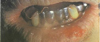
Severe forms of gingivitis, such as ulcerative gingivitis, are observed in severe general diseases of the body. For example, with diabetes or a serious immunodeficiency disease. In this case, the temperature rises, painful ulcers covered with a film appear on the dirty gray gums, and general health worsens.
With gingivitis, the teeth remain stable, since the process does not penetrate deep into the periodontium, the so-called periodontal pocket does not form, and the bone tissue of the tooth socket does not dissolve. Can there be consequences if gingivitis is not treated?
If gums are not treated in time, gingivitis will develop into periodontitis. This is a much more serious and dangerous disease than gingivitis. Often it becomes irreversible, since periodontitis affects and destroys the deep tissues of the periodontium - the ligaments of the tooth and the bone tissue of the jaw.
Periodontal diseases
With mild periodontitis, the symptoms of periodontal disease are mild. Periodic bleeding occurs when brushing teeth or eating hard foods. During the examination, a violation of the integrity of the dental epithelial junction is revealed, and periodontal pockets are present. The teeth are motionless. Due to exposure of the tooth root, hyperesthesia occurs. With moderate periodontitis, severe bleeding is observed, the depth of periodontal pockets is up to 5 mm. The teeth are mobile and react to temperature stimuli. Dental septums are destroyed up to 1/2 the height of the root. With stage 3 inflammatory periodontal disease, patients indicate hyperemia and swelling of the gums. Periodontal pockets reach more than 6 mm. Level 3 tooth mobility is determined. Bone resorption in the affected area exceeds 2/3 of the root height.
With an exacerbation of inflammatory periodontal diseases, a deterioration in general condition, weakness, and fever may occur. With periodontal disease (dystrophic periodontal disease), bone loss occurs. There are no signs of inflammation, the mucosa is dense and pink. Upon examination, multiple wedge-shaped defects are found. Dental cells atrophy gradually. At the initial stage of dystrophic periodontal disease, no unpleasant sensations arise. Patients with moderate severity of periodontal disease experience burning, itching, and hyperesthesia. In severe cases of periodontal disease, due to loss of bone tissue, gaps are formed between the teeth. A fan-shaped divergence of the crowns is observed.
Periodontomas are benign tumor-like and neoplastic diseases of the periodontium. With fibromatosis, dense, painless growths appear without changing the color of the gums. Angiomatous epulis is a mushroom-shaped protrusion of a soft elastic consistency of red color. A separate group includes idiopathic periodontal diseases, accompanied by progressive tissue lysis. Patients develop deep periodontal pockets with purulent discharge. Teeth become mobile and shift.
At the initial stage of Hand-Schüller-Christian disease, hyperplasia of the gingival margin develops. Subsequently, ulcerative surfaces form. Teeth acquire pathological mobility. Purulent exudate is released from periodontal pockets. Papillon-Lefevre syndrome is dyskeratosis of the soles and palms. After the primary teeth erupt, patients with this syndrome experience signs of gingivitis. As a result of progressive periodontolysis, teeth become mobile and pathological pockets appear. Once permanent teeth fall out, bone destruction stops. With Taratynov's disease, bone tissue is gradually replaced by overgrown cells of the reticuloendothelial system with an increased number of eosinophilic leukocytes. It all starts with gingivitis, but soon pathological pockets filled with granulations form. Pathological mobility of teeth is observed.
WHAT FACTORS PROMOTE THE DEVELOPMENT OF PERIODONTITIS?
The causes of periodontitis are the same as those of gingivitis. Often periodontitis is associated with chronic gingivitis. If the traumatic factor persists, microorganisms actively continue to multiply in the damaged gum. Their toxins and enzymes destroy the gums deeper and deeper.
As a result, the connection between the gum and the tooth (the bottom of the periodontal sulcus) is disrupted - a very important protective formation that protects the tooth ligaments and bone from infection. A periodontal pocket appears, and now bacteria, plaque, etc. rush into the depths of the periodontium - this is where periodontitis begins. Next, gradual destruction of the tooth ligaments occurs, and the bone tissue melts.
Periodontitis can be acute or chronic. Typically, acute periodontitis occurs when there is deep, strong trauma to the gums (for example, a long artificial crown, a toothpick). In this case, the periodontal connection may be immediately disrupted - gingivitis and periodontitis occur simultaneously.
Generalized periodontitis is characteristic of serious general diseases of the body - diabetes, other endocrine diseases, radiation sickness, severe diseases of the gastrointestinal tract and cardiovascular system. It is almost impossible to cure such periodontitis without general treatment of the underlying disease.
In the chronic process, pain and swelling are not as pronounced as in acute periodontitis, the amount of tartar and soft plaque increases, the bad breath increases, the gums begin to settle, exposing the neck of the tooth, teeth become sensitive to cold, hot, sour and salty. Tooth mobility appears, unnoticeable at first, but steadily increasing.
Suppuration of periodontal pockets is often observed. Pus is released from under the gum when pressing on the gum edge with a finger. Sometimes, in the absence of outflow of pus, periodontal microabscesses occur - in this case, the patient already requires surgery.
Periodontitis can be one of the causes of some common diseases. The microorganism that causes stomach ulcers is often found in dental plaque. Other bacteria living in dental plaque can lead to the formation of microthrombi (blood clots). Penetrating into the blood (with bleeding gums), they increase the risk of cardiovascular diseases, including myocardial infarction. The entry of microorganisms into the blood can lead to septic endocarditis. There is information about the relationship between chronic dental and periodontal diseases and kidney damage. Remember that the focus of chronic infection in the oral cavity is the entry point for pathogenic bacteria into the body.
Classification of diseases of the oral mucosa
Stomatitis
Stomatitis is an inflammation of the mucous membrane, characteristic of children and adults. Most often, stomatitis is bacterial, viral or fungal in nature. A bad toothbrush with hard, scratchy bristles, poorly fitting braces or crowns, and biting the cheeks and lips can also cause canker sores.
Most often, stomatitis manifests itself in the form of itchy, bright red or whitish sores and erosions on the inner surface of the cheek, tongue or gums. A person may complain of burning and swelling, bad breath, pain when chewing and swallowing. In advanced cases, the temperature may rise, sleep may be disturbed, and the person becomes irritable.
Glossitis
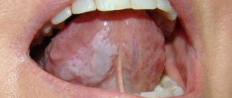
Glossitis is an inflammation of the tongue that can occur either as a result of injury (such as a burn), exposure to pathogens, or as a symptom of certain systemic diseases. Most often, glossitis is manifested by a burning sensation and discomfort in the mouth. The tongue becomes bright red and slightly swollen, and salivation may increase. The patient may complain of loss of taste or changes in the sense of taste, and eating or even just talking causes pain.
Highlit
Haylit (or cheilosis) is a disease in which the lips begin to peel, break, and “sticks” appear in the corners of the mouth. The reasons can be very different: exposure to wind and sun, allergic reaction, chronic diseases with skin lesions (dermatitis, psoriasis, etc.), endocrine pathologies or mycoses.
Oral leukoplakia
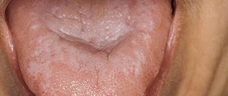
Oral leukoplakia is keratinization of the mucous membrane under the influence of aggressive factors, such as smoking. This condition is considered precancerous and therefore requires mandatory treatment.
Most often, oral leukoplakia appears as whitish, grayish, or red plaques that cannot be removed, rough or keratinized areas, or strange thickenings on the lining of the mouth. As a rule, the patient does not experience pain or discomfort, and therefore does not immediately consult a doctor.
Paradontosis
The periodontium is the complex of tissues that surround the tooth and hold it in place: the gums, periodontal ligament, periodontium, root cementum and bone tissue. Periodontal diseases include: gingivitis, periodontitis and periodontal disease.
Gingivitis
Gingivitis is an inflammation of the gums that most often occurs due to inadequate or irregular oral hygiene. Pathogens accumulate in plaque and tartar, causing inflammation.
With gingivitis, inflammation affects only the surface of the gums and may cause bleeding, swelling of the gums, mild pain or discomfort when pressing, and bad breath. If treatment is not started, the inflammation will go further and affect the periodontium.
Periodontitis and periodontal disease
Very often, patients confuse periodontitis and periodontal disease. Periodontitis is an inflammatory disease of periodontal tissues that causes bleeding gums and leads to the gradual exposure of tooth roots, their mobility and, as a result, their loss. Periodontal disease is a non-inflammatory periodontal disease in which the lining of the gums and jaw bone gradually decrease. Unlike periodontitis, in which tooth tissue is destroyed over several years, periodontal disease progresses very slowly and develops over decades. The patient may not even realize that he has gum disease. Periodontal disease is rare compared to other oral diseases.
DIFFERENCES PERIODONTOSIS FROM PERIODONTITIS
Periodontal disease is a degenerative lesion of all periodontal tissues; this process never occurs in an acute form and is not associated with exposure to bacteria. There is a slow, uniform resorption of the bone tissue of the teeth cells and subsidence of the gums with exposure of the roots of the teeth. Periodontal disease is always a generalized process, that is, all teeth on both jaws are affected. Bone tissue atrophy is usually painless, continuous and, if left untreated, leads to the complete disappearance of the ligamentous apparatus, the walls of the tooth sockets and their loss. Inflammatory phenomena are associated with periodontal disease quite rarely.
The exact cause of the disease is still unknown. It is believed that the onset of the disease is associated with impaired blood circulation in periodontal tissues and endocrine disorders. The development of periodontal disease is promoted by diabetes, cirrhosis of the liver, gastric ulcer, neurogenic diseases, cardiovascular pathology (atherosclerosis), hypovitaminosis and a decrease in the body's overall resistance.
Local factors, for example, the impact of microorganisms on the periodontium, can only aggravate the course of the disease, therefore, in periodontal disease, the primary process is the resorption of bone tissue and the ligamentous apparatus of the tooth, and superficial changes in the gums are secondary.
Periodontal disease progresses slowly. Clinically, it manifests itself with significant changes in the periodontium. This is subsidence of the gums, exposure and increased sensitivity of the necks of the teeth, itching in the gums. Teeth remain stable for quite a long time.
A characteristic sign of periodontal disease is the presence of so-called wedge-shaped tooth defects - damage to the enamel of the teeth near the gums in the form of fairly deep oval cavities. Currently, this phenomenon is explained by a malnutrition of the tooth, inferior enamel and dentin, combined with very strong pressure on the toothbrush during frequent brushing. Periodontal disease is characterized by the absence of periodontal pockets.
Anatomy, physiology
The periodontium
is a complex organ of the oral cavity surrounding the teeth, formed from specialized tissues. It is located on the jaws and its main functions are to support (support) the teeth and their nutrition and innervation.
Nutrition (trophism) is provided by blood and lymphatic vessels that supply oxygen and metabolic products to the periodontal tissue.
There are four main anatomical and functional elements of the periodontium:
- gum,
- periodontium,
- alveolar bone with periosteum,
- root cement
The gum is a soft tissue - a mucous membrane covering the alveolar processes, with an underlying connective tissue layer.
It contains lymphatic vessels (they are involved in regulating fluid pressure in tissues and protecting against microbial agents).
Through the periodontium - the ligament of the tooth, the latter is fixed in the jaw bone.
Periodontium
(otherwise known as desmodont) is a connective tissue formation surrounding the tooth and located between the root cement and the inner cortical plate of the alveolar bone.
This ligament, consisting of the main substance, fibers and cellular elements, like an elastic gasket, absorbs the load on the tooth, giving it minimal mobility. When periodontal disease and death occur, fusion of the bone with the tooth – ankylosis – can occur.
The next element of the periodontium is cement, which is also a hard tissue covering the root of the tooth. It is formed by collagen fibers directed along the root and an adhesive substance.
Bone
The alveolar process, in which the tooth is strengthened by periodontium, is composed of outer and inner dense plates and a spongy part between them.
Spongy bone consists of trabecular septa, the direction and shape of which is determined by the load on the jaws and teeth, and bone marrow.
Bone tissue contains mainly crystals of hydroxylapatite (inorganic) and small amounts of water and organic substances.
The cellular elements of bone - osteoblasts, osteoclasts and osteocytes, under the influence of the nervous and humoral system, determine changes in the shape, volume and quality of this tissue.
The bone tissue of the alveolar process is covered with a dense connective tissue structure - the periosteum; it, with the vessels located in it, plays a significant role in the blood supply to the bone.
The blood supply provides tissues and anatomical formations with organic and mineral substances.
At the level of the enamel-cement junction (neck of the tooth), an anatomical formation stands out in the marginal part of the gum - a vascular cuff, which, like an elastic band, ensures a tight fit of the gum to the tooth due to hydrostatic pressure.
The main source of blood supply to periodontal tissue is the external carotid artery with the maxillary and mandibular arteries branching from it.
Venules and veins collect blood into the internal jugular vein. The periodontium is innervated by the trigeminal nerve and the superior cervical sympathetic ganglion.
Among others, there is an anatomical formation - the dentogingival junction
. This is the connection between the gum epithelium and the neck of the tooth.
It is physicochemical: molecules of epithelial cells adhere to cement structures through gingival fluid cells.
Gingival fluid produced by periodontal tissue plays the role of a protective barrier against microorganisms through the activity of phagocytes and its chemical properties. It should be noted that the periodontium is washed by saliva. It is secreted by the major and minor salivary glands.
In addition to water (99.42%), saliva contains organic substances, salts and trace elements. Organic substances enter the oral fluid from blood serum and are secreted by the salivary glands and microorganisms present in the mouth.
Of the inorganic substances, calcium phosphate and calcium bicarbonate (they take part in the formation of tartar), phosphate and sodium chloride are important.
Diagram 1: periodontium
. a – submucosal layer with lymphatic vessels; b — mucous membrane of the attached gum; c - spongy bone of the alveolar process between the plates of the compact substance; d – periodontal fibers are woven into the root cement; e – gingival margin with vessels of the gingival cuff; e – dentogingival junction; g – tooth enamel; h – dental pulp; and – root cement; j – root dentin.
Age-related changes in periodontium
In the periodontium, with age, there is a decrease in the number of collagen fibers and a decrease in their quality. The epithelial layer of the mucosa becomes thinner, and keratinization is disrupted.
The bone tissue of the alveoli becomes less dense, the cortical layer atrophies.
WHY IS PLAQUE A MAIN CAUSE OF PERIODONTAL DISEASE?
Bacterial plaque is a sticky, colorless film that constantly forms on teeth. If plaque is not removed, it will harden and form a rough, porous growth called tartar. Bacteria in tartar produce toxins (poisons) that irritate the gums, causing them to become red, tender, swollen, and bleeding. As the disease progresses, toxins can lead to periodontal destruction and the formation of pockets that fill with plaque. The bone that supports the teeth is subject to constant destruction. Consistently removing plaque through brushing, flossing, and professional care can minimize the risk of gum disease. However, if no treatment is given, the affected teeth may become loose and eventually fall out.
WHAT IS A PERIODONTAL EXAMINATION?
The history of the disease (anamnesis) is carefully determined. When did it first start, what was done for treatment, do parents and close relatives have this disease, etc. Careful collection of data makes it possible to determine risk factors and sometimes even predict the course of the disease. This is followed by a careful examination of the oral cavity: the frenulum of the lips and tongue, the dental arches as a whole and, if necessary, each tooth separately, as well as fillings, crowns and orthodontic appliances are assessed.
One of the main examination methods is probing the periodontal pocket of each tooth at six points. The presence of tartar and soft plaque, bleeding, suppuration, tooth mobility, etc. is assessed. This labor-intensive analysis is painlessly performed in 15 - 20 minutes using a special electronic probe of the automated computer clinical diagnostic system Florida Probe (USA). The patient’s personal data is stored in the computer’s memory, which makes it possible to predict the course of the process and select treatment, as well as evaluate the dynamics of the disease and the effectiveness of the treatment.
An orthopantomogram is a necessary element in the diagnosis of periodontal diseases. This image shows the teeth, jaw bones, and the partitions between the teeth. Digital orthopantomographs (such as the ones in our clinic) allow you to obtain excellent images with minimal radiation exposure to the patient and evaluate them on a computer screen using auxiliary computer programs.
Principles of treatment of diseases of the oral mucosa
Basic principles of treatment of diseases of the mucous membranes of the mouth, lips and tongue:
- Rational treatment requires contact between the dentist and other dental and non-dental professionals.
- Treatment must be carried out in compliance with the principles of bioethics, these diseases must be considered from the point of view of the state of the whole organism, therefore in most cases one cannot limit oneself to local effects only.
- An axiom for the dentist should be the elimination of all unfavorable irritating factors in the patient’s oral cavity that can support and provoke the development of the pathological process. The use of so-called cauterizing agents and prolonged use of the same mouth rinses is unacceptable.
- Treatment should begin only after at least one preliminary diagnosis has been established and the following requirements have been met: be comprehensive; provide a pathogenetic approach; do not violate the anatomical and physiological characteristics of the oral mucosa; eliminate the pain factor; promote rapid epithelization of lesions; provide for the active involvement of the patient in performing treatment procedures at home.
WHAT IS MEAN BY PERIODONTAL TREATMENT
After collecting all the necessary information, the periodontist will discuss the condition of your gums with you and suggest the most optimal treatment. Most importantly, it must be comprehensive - that is, treatment must cover all identified causes and associated factors that support the disease. Only in this case can it be possible to completely cure or stabilize periodontal disease for a long time.
Treatment will be much more effective if the patient strictly follows all instructions and strictly follows the chosen treatment plan.
Typically, a comprehensive treatment plan consists of general and local treatment and includes all or part of the methods listed below.
Therapy methods
- Etiotropic and pathogenetic therapy aimed at eliminating the cause of the disease (antiviral, antibacterial therapy due to the infectious nature of stomatitis, glossitis, cheilitis, vitamin therapy for hypovitaminosis, treatment of the underlying disease that caused the appearance of a pathological process in the oral cavity) of the mucous membrane;
- Local treatment aimed at eliminating local traumatic factors, the main symptoms of the disease and faster healing of existing erosions and ulcers;
- A general strengthening procedure that stimulates the body's defenses.
ELIMINATING TRAUMATORY FACTORS DURING TREATMENT
Treatment begins with the elimination of traumatic factors - low-quality fillings and crowns with an overhanging or very deeply embedded edge in the gum, bridges, traumatic teeth, etc. are removed.
The so-called selective grinding of teeth is carried out in order to eliminate excessive traumatic load when chewing from overloaded teeth. At the same time, small areas of the teeth are slightly undermined, which do not allow the upper and lower rows of teeth to meet correctly, thereby causing them to be overloaded.
This is an absolutely harmless procedure, since the ground areas of the teeth are polished and coated with fluoride preparations. In this case, caries does not occur.
Professional oral hygiene The next stage of treatment (performed in 100% of cases) is professional teeth cleaning: it allows you to remove even tartar located deep under the gum (this is the strongest traumatic factor). In addition to tartar and soft plaque, dense dark plaque (from smoking, drinking tea, coffee, and other dyes) is simultaneously removed - this allows you to restore the beautiful appearance of your teeth. After removing tartar and plaque, the doctor will definitely polish the cleaned surface of the root and crown of the tooth.
Nowadays, several methods of removing dental plaque are common. Cleaning teeth with a stream of air mixed with cleansing powder and water (the most famous representative is the Air Flow system) perfectly cleanses teeth of soft plaque, dark plaque from tobacco or coffee, even in hard-to-reach places. Ultrasonic scalers are capable of removing tartar of almost any size.
Removing tartar is one of the most important and effective stages in the treatment of periodontal diseases.
Anti-inflammatory therapy Drug therapy of gums allows you to relieve inflammation - cope with pain, swelling, and reduce bleeding gums. Typically, periodontal pockets are washed with antiseptic solutions (chlorhexidine, iodinol, etc.), and various medicinal substances are used (enzymes, antimicrobial, hormonal, anti-inflammatory drugs). Often the gums are covered with a special bandage after the medication is administered.
Self-absorbing Diplen-Dent films impregnated with various medications (antibacterial, improving blood circulation, etc.) are very convenient. Such films are glued by the patient independently to the affected areas of the gums (for example, before going to bed) and, gradually dissolving, release the medicine directly into the gums. In the morning, all you have to do is remove the remaining film from the oral cavity.
Physiotherapeutic treatment There are many methods of physiotherapeutic treatment of periodontal diseases. Laser therapy is most widely used in our clinic. Therapeutic laser radiation has a very wide range of therapeutic effects - relieves pain, improves blood circulation, metabolism, stimulates immune defense.
Gum massage can be performed by a doctor or independently. At home, use your index finger to massage the area of the interdental papilla of the gum with up and down movements (6 to 10 movements per papilla). The massage ends with hygienic rinses. Massage should not be used if the disease is exacerbating or if there are erosions or ulcers on the gums.
Surgical treatment Periodontal surgery is a radical and one of the most effective methods of treating moderate and severe periodontitis. With the help of small operations, it is possible to eliminate periodontal pockets, remove overgrown infected soft tissues, eliminate suppuration of the gums (microabscesses), replant bone tissue or bone substitutes, lift receding gums and close small root exposures.
Every year new developments in the field of surgical treatment, modern materials and treatment methods appear. One of the promising innovations is surgical treatment using cellular technologies, which allows stimulating the restoration of bone tissue. This technology is widely used in our clinic. Surgical treatment is always carried out after removal of dental plaque and therapeutic treatment, when all acute symptoms of gum inflammation subside. All interventions are performed under local anesthesia and rarely take much time. Usually all postoperative phenomena subside after 2 - 3 days, and the sutures are removed, or they dissolve within a week.
The effect of such operations often lasts for a long time. A lot will depend on maintaining oral hygiene, following preventative rules and overall health.
Rational prosthetics and splinting Prosthetics of missing teeth and their permanent splinting (i.e. joining teeth together using a prosthesis or special light-curing threads) are the final stage of the treatment of periodontal diseases. It allows you to fully restore the function of chewing, eliminate aesthetic defects (in the absence of teeth visible when smiling), and unite all the teeth together, as intended by nature.
In this case, the load is distributed evenly throughout the entire dentition, which allows the supporting teeth to function much longer. Even the absence of one tooth causes a number of changes in the bone tissue of the jaw. If, after successful therapeutic and surgical treatment of the gums, rational prosthetics and splinting are not carried out, then the disease may worsen again in the very near future.
Prosthetics are carried out using both removable and fixed dentures, depending on the condition and number of teeth.
After completion of treatment, we advise you to strictly follow all the doctor’s recommendations. Be sure to visit your doctor at least 1-2 times a year for a follow-up examination and professional oral hygiene.
PLANNING TREATMENT MEASURES FOR PERIODONTAL DISEASES
"Medical Information Agency" Moscow 2010
P37 Planning of therapeutic measures for periodontal diseases. - M.: Medical Information Agency LLC, 2010. - 56th: ill.
ISBN 978-5-8948-1835-1
Periodontal diseases are characterized by an extremely high prevalence, a variety of clinical manifestations and often different nature of the course and prognosis depending on a number of local and internal factors. In this regard, planning treatment interventions is a difficult issue not only for young specialists, but also for experienced doctors.
In the presented publication, an attempt was made to create an algorithm for the actions of specialists using both well-known and well-proven means and methods, as well as the latest ones, taking into account the individual characteristics of a particular patient.
For dentists.
UDC 616.314-085 BBK 56.6
© Grudyanov A.I.,
Alexandrovskaya I.Yu., 2010 © Design. Medical Information Agency LLC, 2010
All rights reserved. No part of this book may be reproduced in any form without the written permission of the copyright holders.
Table of contents
Introduction
Chapter 1. Principles of treatment of periodontal diseases
Chapter 2. Initial treatment
Chapter 3. Surgical treatment
Chapter 4. Maintenance therapy.
Literature
Introduction
Treatment of periodontal diseases is one of the most difficult tasks of modern dentistry due to the extreme diversity of clinical manifestations, not only in different patients, but often in one patient with pathology of different parts of the dentition. That is why, despite significant progress in the study of the etiology and pathogenesis of inflammatory periodontal diseases, the strategy of treatment measures, their sequence and effectiveness in each specific case often pose a serious problem, and the proposed standards of treatment turn out to be very conditional.
Nevertheless, in this work we have made an attempt to generalize the accumulated domestic and world experience in systematizing and describing the sequence of implementation of a huge number of means and methods proposed for these purposes in accordance with the taxonomy of periodontal diseases accepted in our country.
Until recently, it was customary to distinguish 3 stages of complex treatment of inflammatory periodontal diseases:
1) initial treatment (pre-therapy);
2) surgical treatment (main treatment);
3) maintenance treatment (maintenance therapy).
In 2003, Peter Fedi in the famous “Periodontal ABC” proposed the fourth stage in the form of restorative therapy (Fedi F. Peter, 2003), and in 2008, Herbert Wolf proposed the “zero stage” as an independent one - emergency treatment (Wolf F. Herbert, 2008).
Chapter 1
HOW MUCH WILL THE TREATMENT COST?
The cost of periodontal treatment will depend on the type and amount of work planned. When considering an investment in your own health, consider that treating gum disease is cheaper and better for your health than restoring a tooth lost due to untreated periodontal disease.
New direction – aesthetic surgical periodontology
Recently, such a direction as aesthetic surgical periodontics has appeared and is actively used in surgical periodontology. She deals with the correction of changes in the position of the gingival margin relative to the neck of the tooth, and gum growths. Aesthetic surgical periodontics includes a large number of new surgical techniques, depending on the clinical situation and the desired aesthetic effect.
ARE THERE METHODS FOR PREVENTION OF PERIODONTAL DISEASES?
- Prevention of periodontal disease consists of several simple points:
- Brush your teeth 2 times (morning and evening) after meals. Use a toothpaste recommended by your doctor to protect your gums. Use an electric toothbrush - it perfectly cleans your teeth and massages your gums.
- Do professional oral hygiene at least 2 times a year, and at the same time you can undergo an examination by a periodontist. Do not use any medications for the treatment or prevention of periodontal disease (gels, ointments, tablets, etc.) without consulting your dentist.
- Do not eat only soft and delicate foods - your teeth should receive normal, natural stress.
- Your diet should be well balanced, contain the required amount of proteins, fats, carbohydrates, and vitamins. If you are deficient in vitamins, take multivitamin complexes.
- Lead a healthy lifestyle.