Today, many patients when visiting dentistry complain about the loss of the natural shade of tooth enamel. Why does enamel become dull and lose its natural color? There are actually a huge number of reasons, and it is to them that we will pay attention today, but a little further. Now we note that this problem can be dealt with using modern teeth whitening technologies, Zoom whitening, Brite Smile, Opalescence, which are successfully used in medical science.
Changing the color shade of tooth enamel: varieties
There are two types of loss of natural tooth enamel color:
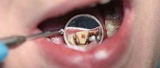
- Internal change. Occurs in the inner part of the tooth structure - dentin. The tooth becomes covered with gray, yellow spots or darkens.
- External change. The process takes place in the outer layer; deterioration is indicated by the appearance of yellow spots, white stripes, and dips on the surface of the tooth.
It is worth noting that external discoloration can be treated with special whitening technologies. Internal change is more difficult to cure. Its treatment may require aesthetic methods, such as veneers.
Medical Internet conferences
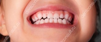
Relevance. The term “dental discoloration” means a change in the color of the crowns of natural teeth. Changes in tooth color are a fairly common cause of impaired smile aesthetics; the prevalence of this condition in the structure of dental pathology is about 15% [1]. A beautiful smile reflects a person’s social success, his health and simply predisposes people to him, but unfortunately, the rhythm of life of a modern person does not always allow him to maintain the natural appearance of his teeth and their health. A large number of bad habits, in particular smoking, poor oral hygiene, eating a large number of foods with a high content of coloring pigments - all this negatively affects the color of teeth. People have to pay a lot of money to try to change the current situation with discloritis of their teeth by visiting the dentist’s office, where they undergo procedures in the form of professional whitening, professional oral hygiene, which are not always pleasant and take up a large amount of their personal time. Therefore, it is an important value to realize in time what influences the color of teeth to a greater extent in everyday life and try to avoid these negative factors
Purpose: structuring the etiological factors of dental discoloration and identifying the relationship between the causes of discoloration and shades of teeth.
Tasks:
1. Display the structure of etiological factors for changing the color of teeth with a description of the mechanism of action of each factor.
2. Establish the relationship between the most common etiological factors of discoloration and tooth tones.
Materials and methods. An analysis of scientific articles and a review of modern educational literature on the topic “changes in tooth color” were carried out. A survey was conducted of 30 students of the Faculty of Dentistry of the Federal State Budgetary Educational Institution of Higher Education "Saratov State Medical University" named after. IN AND. Razumovsky" of the Ministry of Health of the Russian Federation aged from 20 to 25 years and 30 schoolchildren of the MBOU "Secondary school No. 33 named after P.A. Stolypin" in the city of Engels, aged from 13 to 17 years (the average age of those examined in both groups was 17.8 years). Of these, the number of males is 27 (45%), females 33 (55%). In these groups, a survey was conducted, where respondents were asked to select the most common reasons associated with changes in the color of their teeth. Then the color of the teeth was assessed using the Vita color scale. Further statistical processing was carried out manually.
Results and discussion. In the structure of etiological factors of dental discoloration, it is traditional to distinguish two main directions - temporary and permanent color changes.
Temporary changes in tooth color. This group includes those discolorations that, as the name suggests, can either intensify or disappear over time, depending on the strength of the etiological factor [2]. This type of discoloration includes the very common ones today - changes in the color of teeth due to the influence of food dyes contained in large quantities in foods and drinks, which are available in a wide range in any grocery store and chain catering establishment. When it comes to drinks, coffee comes to the fore, a drink made from roasted plant seeds. By consuming it hot, a person creates conditions for staining his teeth, because in the interval between the expansion and contraction of the surface layers of enamel, which occurs as a result of temperature fluctuations, the penetration of coloring substances into the tooth occurs. As a result, people who abuse coffee develop a light brown or yellow tint to their enamel over time. Tea containing the coloring pigment tannin has a similar mechanism of action. It should be noted that discolorations can be of two types: internal and external. If the coloring substances do not penetrate directly into the tooth structure, but only settle on its surface, then this color change will be external. If the opposite happens, and the pigments penetrate the enamel, dentin and cementum of the tooth, then such staining will be called internal. It follows that even when drinking non-hot coffee and tea, coloring pigments can settle on the surface of the enamel and cause staining of the tooth if a person does not rinse the mouth with water after drinking the drink [3].
Colored highly carbonated drinks, which have become companions of modern fast food, and which are often consumed by people at a young age who constitute a risk group, can also cause tooth discoloration. The presence of an acidity regulator in the form of citric acid (food additive E330) in these drinks negatively affects the health and structure of the teeth, and the carbohydrates and sugars contained in large quantities (about 20% in total) are a substrate for reproduction and contamination of the oral cavity cariogenic microflora, which will subsequently lead to demeniralization processes and damage to the integrity of the tooth. In the future, as described earlier, the pigments will penetrate into the internal structures of the tooth and cause staining. In ongoing scientific studies, situations involving the impact of aggressive highly carbonated drinks on extracted intact teeth were simulated, where they were placed, after cleaning and disinfection, in a container with various liquids for a period of one day. As a result of the experiment, it was found that the most aggressive liquid was the Coca-Cola drink, which caused staining of the enamel to a depth of 0.81 mm [4].
When talking about products that have the strongest coloring properties, it is worth focusing on sweets and berries. For example, chocolate, unlike previous products, does not cause temperature fluctuations in the oral cavity and does not contain aggressive acids, but having greater viscosity and adhesion, it contacts the tooth surface for a longer period of time, facilitating the penetration of pigments into the surface layers of enamel , and the formation of dental plaque. And we should not forget that even such healthy foods as berries can harm the health and beauty of teeth if their consumption is not controlled. Containing both a large amount of pigment substances and enamel-damaging acids, they also contribute to permanent discoloration of teeth. Raspberries, grapes, blueberries and currants are considered the most “unfavorable” for teeth.
Also included in this group are unstable dental discolorations, which occur as a result of a person’s bad habits. The most common habit that leads to yellowing of teeth is smoking. The determining factor in mechanical damage to teeth when smoking is the huge temperature changes that occur in the oral cavity while smoking a cigarette. According to research, temperature fluctuations reach 10-15 times the norm [5]. As a result, the slightest microcracks appear on the surface of the enamel, through which pigment particles will penetrate deep into the tooth and cause staining.
The next etiological factor from the group of temporary discolorations is unsatisfactory oral hygiene. Due to non-compliance with the rules of brushing teeth, and in particular: brushing less than twice a day, the movements of the toothbrush do not correspond to the methods of brushing teeth, insufficient time devoted to brushing, untimely change of the toothbrush, weak force applied to the toothbrush – over time, all these factors will contribute to the accumulation of plaque of varying degrees of pigmentation on the teeth [6]. According to its species structure, plaque comes in two main types – green and brown. The first type is most often found in young people. Located in a thin layer on the surface of the frontal group of teeth, it contains chromogenic microflora (fungus Lichen dentalis), which determines its color. The brown type can most often be found in smokers, people who abuse tea and coffee drinks, as well as people working in enterprises associated with the production of brass and bronze products, where there is a large amount of suspensions of these materials in the air, which are deposited on the surface over time teeth.
The next direction in the structure of etiological factors of tooth dislorites is permanent staining. In turn, it is divided into permanent congenital color changes and permanent acquired ones.
Permanent congenital color change. The first cause considered in this subgroup will be “tetracycline teeth.” This disease is rarely encountered in the clinical practice of dentists, due to the increased role of preventive work in antenatal clinics, but is still often mentioned in textbooks and literature, so it makes sense to consider it in more detail. Changes in tooth coloring are associated with the mother taking antibiotics from the tetracycline group during pregnancy (starting from the fifth month of intrauterine development), as well as treatment of a child under 7 years of age with the same drugs. The follicular development of teeth that occurs at this time is disrupted; tetracycline binds to hydroxyapatite of hard dental tissues using chelate compounds to form a tetracycline orthophosphate complex, which in the future will be responsible for the lemon tint of teeth. After some time, as a result of food pigments and the photochemical reaction that occurs due to sunlight, the color of the teeth will turn dirty brown. The possibility of the appearance of “tetracycline teeth” remains in the adult population taking these medications, in which the penetration of the antibiotic into the internal structures of the tooth will be due to the diffusion of the drug from saliva [7].
A pathology such as hemolytic diathesis, which develops as a result of incompatibility between the blood of mother and fetus, can also lead to the development of congenital discoloration. As a result of the Rh conflict, hemolysis of the erythrocytes of the newborn and fetus occurs with the formation of a large amount of unconjugated bilirubin. Penetrating and accumulating in the structure of temporary teeth (mainly in dentin), over time, bilirubin breaks down, which leads to a change in the color of temporary teeth, ranging from yellow to brown or greenish-blue shades.
With congenital malformations of the biliary system, when, due to various etiological reasons, the outflow of bile is disrupted and its stagnation in the bile ducts, a disruption of the connection between the blood and bile capillaries occurs. As a result, this leads to an increase in the level of direct and indirect bilirubin and the further mechanism of color change is similar to that described in hemolytic diathesis.
In Gunther's disease (synonyms: congenital porphyria, erythropoietic uroporphyria), transmitted in an autosomal recessive manner, uroporphinogen-3-cosynthase deficiency occurs, which leads to excessive formation of intermediate products of heme synthesis, namely uropophrinogen and coproporphinogen. Subsequently, they are deposited in the hard tissues of the teeth, giving them a purple-brown color, and fluoresce in ultraviolet radiation with either an orange or a red tint [8].
In fluorosis, excess fluorine enters the enameloblasts during enameloblasts and forms a strong hydroxyfluorapatite structure. Subsequently, the excess amount of fluoride is fixed on the enamel surface in the form of a calcium fluoride compound, covering the enamel hydroxyapatite. Having different optical characteristics, calcium fluoride changes the color of the enamel surface to a more matte one. The further mechanism of color change during fluorosis is not fully understood, but there are suggestions that areas that become darker over time are softer, where dyes penetrate to a greater extent in the future [9].
The change in color during systemic hypoplasia to whiter is explained by impaired development of tooth tissue and demineralization processes. The cause of local hypoplasia can be injury to a baby tooth during the time period when the formation of a permanent tooth germ occurs. In this case, the breakdown products of blood cells enter the tissues of the permanent tooth germ, which are included in the enamel matrix of the permanent tooth during its mineralization, changing its color to gray.
Permanent acquired color changes. Age-related change in the color of teeth is a polyethylological phenomenon and only with the combination of a large number of factors, over time, in older people, a change in the coloring of the crowns of teeth occurs. Firstly, over time, the quantitative ratio of tooth structures changes - the amount of more transparent enamel decreases and the percentage of replacement dentin increases, which in turn is not only darker in shade, but is also more easily susceptible to pigmentation, since it has greater permeability. In addition, with age, the pulp chamber decreases, which also leads to a decrease in the transparency of the tooth. Secondly, an important role is played by the combination of exogenous influences that were already described earlier in the article - smoking, the presence of food pigmentation, the appearance of dental plaque, microcracks and defects in the surfaces of teeth. All this together forms the appearance of an “old” tooth over time.
The color of teeth can also change as a result of the carious process. Already at the initial stages of the pathological process, as a result of subsurface demineralization, a change in the color of the teeth occurs - they become chalky. In the future, over time and the progression of the process, the superficial and deep layers of enamel and dentin will be saturated with pigments diffusing from the oral fluid. At later stages of the process, the staining of necrotic dentin black will occur due to the microflora of the oral cavity, and in particular, changes will be caused by Porphyromonas endodontalis and bacteria of the genus Prevotella.
The crown may turn pink as a result of an injury. This occurs as a result of a complete rupture of the neurovascular bundle in the area of the apical foramen. Over time, due to the decomposition of hemoglobin and the appearance of hemosiderin and other pigments of decomposed blood in the pulp chamber, the color of the tooth will change to darker shades - burgundy and even blue-black. When the traumatic factor is insignificant, and when the structure of the neurovascular bundle is not disturbed, the color of the crown may change to more yellow shades - this is a consequence of the deposition of tertiary dentin and a decrease in the size of the pulp chamber [10].
Discolors during tooth depulpation have a similar mechanism of action for color changes. The areas of pulp under the hanging walls of dentin that have been preserved after poor-quality preparation will also decompose into iron-containing complexes with the subsequent coloring of the teeth in a dark sulfur color.
Violation of the technique of filling root canals, when, due to incorrect actions of the doctor, the material ends up in the coronal part of the tooth, in turn, can also lead to dental discloritis. This is true for pastes containing eugenol, which lead to a yellow-brown tint of teeth, for materials containing formaldehyde (resodent, resorcinol-formalin paste, forfenan), which change the color of teeth to pink. Also, if a mistake is made during endodontic procedures, when a section of a broken metal instrument is left in the root canal, the teeth will change color to a dark gray color due to diffusing metal ions.
When conducting a survey of students and schoolchildren, where they were asked to select the causes of discoloration they had, reflected in the article, the most common factors were identified - poor oral hygiene, consumption of drinks containing a large amount of coloring pigments, consumption of coloring products and the presence of signs of carious lesions in the oral cavity. Discolorations due to poor quality treatment and nicotine pigmentation were less common. The structure of identified predisposing factors for dental discoloration among those examined is presented in the table.
Structure of discoloration factors (n=60)
| Factors | Students (20-25 years old) (n=30) | Schoolchildren (13-17 years old) (n=30) | ||
| Abs. | % of all students | Abs | % of all schoolchildren | |
| Consumption of foods with high coloring power | 13 | 43 | 28 | 93 |
| Drinking drinks with high coloring power | 13 | 43 | 18 | 60 |
| Poor oral hygiene | 11 | 36 | 17 | 56 |
| Dental caries | 20 | 66 | 10 | 33 |
| Discoloration due to poor treatment | 3 | 10 | 3 | 10 |
| Nicotine pigmentation | 3 | 10 | 0 | 0 |
Based on the data in the table, we can conclude that in the group of schoolchildren examined, the most common factors of dental discoloration are food pigmentation - consumption of both foods and drinks containing a large amount of coloring pigments, as well as poor oral hygiene. For a group of students, dental caries comes out to be the first predisposing factor. It should also be noted that in the group of students such a bad habit as smoking appears that contributes to discoloration of teeth. This leads to recommendations aimed at eliminating discolouration. Persons who abuse the consumption of coloring foods or drinks must observe hygienic measures: after eating such food, you must thoroughly rinse your mouth with water, or, if possible, brush your teeth. Persons with unsatisfactory oral hygiene need to master the standard method of brushing their teeth under the supervision of a dentist, brush their teeth for 3 minutes 2 times a day - in the morning before breakfast and in the evening before bed, use additional hygiene products (floss, toothpicks, interdental brushes) for cleaning contact surfaces, change your toothbrush to a new one every three months. Persons with carious lesions are recommended to sanitation of the oral cavity, and the only measure to prevent nicotine pigmentation is to give up this bad habit.
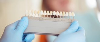
Next, the tone of the teeth was assessed using a color scale. The group of examined patients had tooth colors from A1 to B4, the most common were the following: A2 (18% of those examined) and D4 (13% of those examined), C2 (13% of those examined). During further statistical processing, the examined patients were divided into groups corresponding to their teeth tone, and the influence of each etiological factor presented in the table on each group was assessed. During this process, the clearest relationship was established between poor oral hygiene and discoloration of their teeth. Students with teeth shade A1 had an insufficient level of hygiene in only 25% of cases, while in the following groups with darker shades this percentage increased (B2 - 50%, D2 - 66%, A2 - 50%, C1 - 50%, C2 – 50%, A3 – 66%, D3 – 100%, B3 – 100%). The same trend was observed in the group of schoolchildren (D2 - 0%, A2 - 0%, C1 - 33%, C2 - 62.5%, D4 - 50%, A3 - 100%, D3 - 100%, B3%, A3 .5 – 100%, B4 – 100%). In the case of other etiological factors, such a clear relationship was not established. For example, the consumption of coloring drinks in the group with a light shade of B2 was 100% of those surveyed, while in the group with a tooth shade of B3, which is much darker, it was only 50%. The presence of a small number of smokers among those examined did not allow us to evaluate this parameter, but it should be noted that the lightest shade of a smoker’s teeth was C1, which is a very dark shade, allowing us to say that smoking, as well as poor hygiene, have a direct relationship with color change teeth.
Conclusions:
1. In the group of examined schoolchildren, the most common factors of dental discoloration are food pigmentation - consumption of both foods (93%) and drinks (60%) containing a large amount of coloring pigments, as well as poor oral hygiene (56%). For a group of students, dental caries comes out on top as a predisposing factor (66%). It should also be noted that in the group of students such a bad habit as smoking appears (10%).
2. As a result of the study, after statistical processing, the clearest relationship was revealed between poor oral hygiene and developing dental discoloration. Students with teeth shade A1 had an insufficient level of hygiene in only 25% of cases, while in the following groups with darker shades this percentage increased (B2 - 50%, D2 - 66%, A2 - 50%, C1 - 50%, C2 – 50%, A3 – 66%, D3 – 100%, B3 – 100%). The same trend was observed in the group of schoolchildren (D2 - 0%, A2 - 0%, C1 - 33%, C2 - 62.5%, D4 - 50%, A3 - 100%, D3 - 100%, B3%, A3 .5 – 100%, B4 – 100%). At the same time, the establishment of the same clear relationship among other etiological factors did not occur, which indicates their lesser role in the occurrence of changes in tooth color.
The main reasons for changes in the shade of tooth enamel
- Consumption of food and drinks. Internal spoilage can occur from eating a wide variety of foods if proper oral care is not maintained.
- Tobacco use causes damage to the outer layer of tooth enamel.
- Inadequate dental care. If you do not brush your teeth regularly or with low-quality toothpastes, there is a risk of discoloration of the enamel.
- Use of medications. Any medications for high blood pressure can affect your dental health. If a pregnant woman uses tetracycline antibiotics, there is a risk of loss of color of the enamel and the newborn.
- Diseases. There are some infections in pregnant women that can affect the color of the newborn's tooth enamel, and teeth can also lose their natural color in case of dentin disease.
- The presence of dentures in the case of using silver amalgam during care can give the enamel a grayish tint.
- Genetic predisposition.
- Aging. As you age, tooth enamel may become yellowish. Also, soft or hard plaque may form on the surface of the teeth, which also causes discoloration of the teeth.
- Medical procedures. There are procedures, for example, chemotherapy, in which one of the side effects is the effect on the color of tooth enamel.
Most often, a person can cope with the problem, especially if it is caused by an external discoloration. All that is required is regular visits to the dental office and proper hygiene using quality toothpastes.
Immediately after eating food or drinks (red wine, tea, coffee), it is recommended to immediately rinse your teeth with water, but it is better to brush your teeth with toothpaste. After smoking, it is also necessary to brush your teeth, otherwise an unpleasant yellowish tint may appear on the tooth enamel in the near future.
Methods to combat tooth staining
Today, there are many options for teeth whitening in dental practice. Patients often try to whiten enamel at home, using cheap and very aggressive means - citric acid, activated carbon, hydrogen peroxide solution. The specialists at Dr. Granov’s dental clinic strongly do not recommend that you engage in such “amateur activities.” The maximum you can achieve using these methods is a slight lightening of 0.5-1 tone, and only if you regularly use the recipe. In this case, your enamel will suffer catastrophically - it will become thin, fragile and hypersensitive. The only acceptable method of dealing with teeth staining at home is the use of professional solutions and whitening toothpastes. Consult a specialist regarding the selection of such a product. In our clinic you can access the following services:
- Whitening “Air Flow”;
- Whitening "ClinPro";
- Teeth whitening “Opalescence”;
- Whitening “ZOOM3”;
- Whitening using laser equipment.
We also provide a professional ultrasonic cleaning service, which can make your teeth 1-2 shades lighter, returning them to their natural whiteness. Sometimes this is enough to achieve results. Our specialists offer their patients the service of selecting home whitening products (trays and solutions). In some cases, non-invasive whitening is not enough, and the dentist may offer you artistic restoration methods (veneering, lumineering, etc.). If you want to find out the reason for the staining of your teeth, or want to get rid of the problem of yellowing of the enamel, we invite you to an in-person consultation at our clinic.
Dental abnormalities that may cause discoloration of tooth enamel
Changes in tooth color can occur as a result of changes in their size or shape. The color of the tooth may change depending on the number of teeth and the period of eruption.
Complete edentia is extremely rare, but the absence of several teeth is a common pathology. It is explained by endocrine disorders, which can cause a delay in teething and, as a result, a change in color.
With tuberculosis, neurological diseases, rickets, retention of the molars, as well as the upper canines (permanent), occurs.
In diseases associated with osteogenesis, patients most often have small teeth and a bluish tint to the tooth enamel. It is worth noting that the teeth of people with such diseases wear out quickly and are especially susceptible to disease. Pathologies in the development of dental dentin and tooth enamel cause amber, opal, bluish and brown tints of teeth and insufficient size.
Endogenous (internal) factors
Internal causes of teeth staining are more complex and multifaceted than external ones. It should be understood that under certain conditions these can occur in childhood. For example, if a child consumes too much fluoride during a change in bite, his teeth may acquire a certain shade, regardless of hereditary or national characteristics. From the clinical practice of our specialists, it is clear that enamel that has undergone changes due to internal factors whitens worse. Often, to achieve optimal results, it is necessary to eliminate the root cause of the problem. Endogenous factors for tooth staining:
- Disturbances in the development of dentin and enamel in childhood and adolescence (fluorosis);
- Metabolic disorders (erythropoietic porphyria, exposure to bilirubin, etc.);
- Schematic use of antibiotics of the tetracycline group;
- Root resorption;
- Age-related changes in enamel.
With age, teeth can also change their shade. This occurs against the background of thinning enamel in older people. At the same time, secondary dentin is formed in the internal structures of their teeth, which aggravates the situation. The enamel loses its transparency, causing the teeth to look dark and matte, which is called the “senile smile.” Sometimes tooth staining is associated with internal chronic pathologies of a systemic type, especially if they accompany the patient for most of his life. An equally common trigger for the violation of the natural whiteness of the enamel in this case is constant treatment. We must not forget that some medications negatively affect dental tissue. And if you take them constantly, you can achieve a permanent deterioration in the aesthetic qualities of your smile.
How to restore the color of tooth enamel?
In many cases, with the exception of diseases and pathologies, patients have every chance to influence and change the color of tooth enamel for the better, without even going to the dentist. The surest way is to eliminate all foods and drinks that contain food colorings.
In difficult cases, various modern technologies, professional teeth cleanings help restore whiteness, and in the most difficult cases, prosthetics are performed.
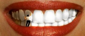
There are many ways to restore the natural color of enamel; by contacting our center, each patient can count only on quality, namely the complete restoration of tooth enamel.
How to keep your teeth white
To keep your teeth white, you need to:
- Perform good oral hygiene, including brushing, flossing, and using mouth rinse
- Using a waterpik at least twice a week to get rid of plaque
- Once or twice a year, have your mouth professionally cleaned by a dental hygienist to remove tartar.
- Limit or eliminate smoking, strong tea and coffee
It is also important to make regular visits to the dentist for diagnostic procedures. This will allow timely identification of problems and identification of deterioration of the enamel.
Symptoms accompanying tooth darkening
If the cause of the defect is a bruise, then the symptoms will be pronounced. More often, the impact damages the upper or lower front teeth. At the same time, the baby feels pain, which only increases over time. Pain is especially evident when pressing on a sore spot or chewing food. In addition to pain and staining, other symptoms may occur:
- poor tooth mobility;
- swelling of the gums;
- hyperemia;
- deformation of individual areas of the mucosa;
- formation of hematomas;
- increase in temperature (in some cases).
If the blow to the jaw was strong, the ligaments at the site of the injury may be torn, the joint may be damaged, or the alveolar process may be fractured. When trying to open or close the jaw, the child will experience pain.
Treatment of a darkened tooth
Darkening of the tooth near the gum requires immediate consultation with a doctor, regardless of the cause. If you leave the disease to its own devices, you can face more dangerous and serious consequences.
The doctors at our regional dental clinic are ready to help. We use modern equipment, progressive therapeutic practices and always put the patient's comfort as a priority. Therefore, treatment of a darkened tooth will be quick and effective. Don’t let the disease lead to dangerous consequences - make an appointment with the dentist!
What is the treatment
If the darkening of dental tissues is associated with dental diseases, the doctor will act according to the protocol. It is important to eliminate negative factors and carry out correct treatment. In case of caries, the dentist will remove the damaged tissue and install a filling. If the disease is advanced, endodontic treatment may be necessary, in which pulp removal is performed and the canals are filled. When pigmentation is caused by a general disease of the body, to eliminate it you need to cure the disease itself.
It is worth considering the situation in more detail if the tooth has darkened after an impact. What to do in this case? It all depends on the force of the impact and damage. If the bruise is not severe, most often only medical supervision is required (even up to 2-3 months) without any treatment. The tooth is provided with rest, excluding chewing and pressure on it. You need to remove solid food from your child’s diet for several days. To remove the load from baby teeth, their cutting edge is ground off. This technique is not used for indigenous people.
If the bruise is severe, the first thing you can do is give your child painkillers. The doctor’s actions depend on the clinical picture. If the pulp is damaged, they are gradual:
- tooth opening and pulp removal are carried out;
- the dental cavity is thoroughly disinfected;
- The channels are tightly filled with filling material.
If a permanent tooth has darkened as a result of a bruise, you can whiten it. After the procedure, the enamel will acquire a more or less natural color. To quickly restore oral health after injury, the dentist additionally prescribes physiotherapy and anti-inflammatory drugs.
Complete dislocation requires tooth extraction, after which the child’s condition returns to normal. In some cases, they may try to save the integrity of the dentition. To do this, the tooth is splinted using ligatures, caps, staples and other structures. The last steps are possible if the roots are sufficiently developed. Children aged 2–3 years often have to undergo removal.
Consequences of a tooth bruise
Darkening of the dentition provokes psychological discomfort, especially if these are the front teeth. Any injury does not go away without a trace. The tooth can be dislocated, cracked, broken, or sometimes embedded in the gum. Even if at the time of treatment the determination of the vital activity of the pulp gave a positive result, after the end of therapy it can still gradually die. This process is accompanied by damage to nerve endings, which provokes inflammation. More serious complications are also possible:
- cyst formation;
- manifestation of periodontitis;
- stopping root development.
These unpleasant consequences are possible in both baby and permanent teeth. Advanced pathology can even lead to tooth loss.
The rudiment of a permanent tooth often suffers from injury to a baby tooth. His enamel may develop poorly, and the risk of hypoplasia formation increases. The rudiment sometimes even dies.
How to diagnose
At the diagnostic stage, it is important to determine why the tooth has darkened. The doctor will perform palpation and visual examination. But in order to make an accurate diagnosis, assess the degree of damage or carious lesion, one cannot do without radiography. Based on its results, the doctor determines what to do to eliminate the problem. In some cases, consultation with other specialists, such as a gastroenterologist or endocrinologist, may be necessary.
X-rays can also be used to rule out a fracture of the root or alveolar process due to a bruise. If the periodontal area is damaged, the defect will definitely be visible on the image. After the impact, the dentist should monitor the condition of the pulp for several days. For this purpose, electroodontodiagnostic methods are used. If even initial signs of death are detected, the affected areas will be removed so that an inflammatory process does not develop.
Features of baby teeth
The first teeth of babies have porous, thin enamel, which cannot effectively resist the attack of harmful microorganisms. The pulp cavity is wide. Once bacteria gets inside, they spread quickly. If a child has a weakened immune system, the pathogenic process affects the “neighbors.” Carious lesions are possible even in a one-year-old baby. We are talking about the so-called “bottle” caries, when the baby falls asleep while feeding. The sweet mixture remaining in the mouth creates favorable conditions for the development of pathogenic microflora.
Since caries in children progresses quickly, after some time the dentin is damaged. A carious cavity forms inside. But there is not always a visible stain on top. Blackening of the tooth from the inside may occur. Various factors contribute to the development of caries:
- insufficient hygiene is a common occurrence in children;
- genetic predisposition;
- poor nutrition, including excessive consumption of sweets;
- the influence of negative factors during the formation of teeth in the embryo - previous illnesses of the pregnant woman, taking certain medications.
Saliva has antibacterial properties and helps neutralize bacteria. In children this function is not as pronounced as in adults. She is also depressed by non-compliance with hygiene rules and a large amount of sweets in the diet.