How to identify milk caries
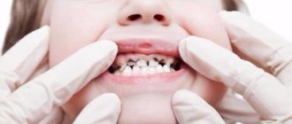
It is quite simple to identify caries in young and older children. White or brown spots appear on the teeth, a painful reaction to hot and cold is observed, and the child may develop bad breath. When these first symptoms of caries appear, it is already worth sounding the alarm, since the carious process is developing very rapidly. It is characterized by almost instantaneous damage to several teeth, and if measures are not taken, the entire dentition may be affected. Of course, it is often difficult for a child to articulate that his baby tooth hurts. He may refuse to eat or chew on only one side. This should also alert parents and encourage them to take their child to pediatric dentistry.
What to do
Treatment methods for dental caries in children depend on the stage of development of the defect. They are invasive and non-invasive. The latter are used at the initial and superficial stages. Non-invasive methods include:
- remineralizing therapy (saturation of enamel with phosphorus, magnesium, calcium and other useful elements);
- fluoridation (coating enamel with fluorine to prevent demineralization);
- ozone therapy (ozone treatment to destroy pathogenic bacteria);
- silvering (applying a solution of silver nitrate to the affected areas).
These procedures are painless, take 5-15 minutes, and are suitable for children under 3-5 years of age.
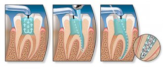
Deep caries requires preparation of the carious cavity followed by filling it. The invasive method is used to eliminate large defects. To remove destroyed tissue, the dentist uses a drill and a stream of water. The cleaned cavity is dried and disinfected.
After sanitation, the carious cavity is filled with filling material. It is given the anatomical shape of a dental crown, then polished so that the child does not feel discomfort. If the enamel is weak, special pastes and preparations are used to strengthen it.
If there is significant tooth decay, when conservative measures are not able to save it, they resort to extraction. Then the missing dental unit is artificially restored so as not to disrupt the formation of the child’s bite. If you consult a dentist in a timely manner, it is possible to prevent the spread of caries to neighboring teeth and prevent serious complications.
Treatment for caries of permanent teeth in children is the same as in adults. For filling, composite compounds are used that follow the contours of the carious cavity.
Where does childhood caries begin?
There are more reasons for this than meets the eye. This includes improper oral hygiene, sharing cutlery with adults who have caries, and much more. However, the main causes of occurrence are the influence of consumed carbohydrates that destroy the tooth, accidental infection from a carrier of a carious infection, a genetic factor, the immaturity of the children's skeletal system, and improper use of pacifiers and nipples.
- Of course, the main and most common reason why childhood caries occurs is poor oral hygiene and the influence of sweets. According to statistics, caries develops in young children in 73% of cases. Sucrose, glucose and fructose are the acids that are formed during the fermentation of carbohydrates and lead to the destruction of enamel. Immediately after ingesting carbohydrates, the pH of saliva decreases from 6 to 4 and pathogenic bacteria settle in food debris that is not cleaned from the teeth.
- Remember the children's saying “from mouth to mouth you get a germ”? It turns out that she is not so far from the truth. The fact is that caries is an infection, an infection. That is, a loving parent, being a potential carrier of carious microbes, does not even suspect that by kissing his baby or sharing dinner with him, he can personally infect his child!
- Another reason why caries may occur on baby teeth is genetic. Teeth begin to form during the prenatal period, in the first trimester of pregnancy. Therefore, smoking by a negligent parent, her illnesses suffered during this period, or taking medications can lead to disruption of the proper development of the child’s teeth.
- Caries in children 2 years old, that is, early caries in children, may be associated with the following factor: baby teeth have a low degree of mineralization and erupt “immature”, and only then “mature” in the oral cavity. Final mineralization lasts for one and a half to two years for baby teeth and about three years for permanent ones. Pediatric dentists call such periods of “maturation” the most susceptible to caries. Therefore, the reasons why caries occurs in a child of 2 years and at an earlier age may also be chronic diseases, the effects of medications, the composition of saliva and the fluoride content in water and food.
- Another reason for the development of caries in very tiny children is the incorrect use of pacifiers. A baby who falls asleep with a bottle in his mouth runs the risk of getting caries on his front teeth, so-called bottle caries. In this case, prolonged contact of the sweet liquid with the teeth leads to caries of all front teeth. In this case, the disease spreads around the entire visible part of the tooth along the perimeter.
As you can see, there are more than enough reasons for the appearance of caries. But they all boil down, as a rule, to demineralization of teeth and destruction of hard tissues. Characteristic changes occurring in teeth can only be diagnosed by a dentist. Further treatment will depend on how severe the caries has developed. What are the stages of development of caries in primary teeth?
Therapy methods
Treatment of caries detected at the initial stage of development is carried out conservatively using methods of silvering or deep fluoridation of enamel. When carious cavities are detected, they are cleaned (using a drill or chemical preparation) and filled with quickly hardening composite materials.
All therapeutic procedures are performed only after the anesthesia procedure. In this case, preference is given to local anesthetics, but if necessary, treatment can be carried out under general anesthesia.
Stages of development of childhood caries
Dentists divide caries of primary teeth into several types according to the depth of damage:
- elementary;
- surface;
- average;
- deep.
Initial caries
Initial caries can be recognized as follows: white spots of various shapes and sizes appear on the enamel, but there is no pain. In advanced cases, initial caries progresses - the spots darken, becoming brown and then black.
Superficial caries
With superficial caries, the defect in the dental tissue is located within the enamel, and the carious cavity can be either light or dark. However, in this case, pain occurs when the tooth comes into contact with sweet, sour or salty foods. In this case, treatment of the baby tooth and filling of the cavity is already required.
Average caries
Medium caries affects the enamel and inner tissue of the tooth (dentin). In addition to the unpleasant sensations from sweet, sour and salty, pain from hot and cold is added. In this case, as with superficial caries, filling is necessary.
Deep caries
With deep caries, the enamel and a significant part of the dentin are affected. If treatment is not carried out at this stage, then caries can affect the pulp of the tooth and then reach the root, often resulting in a cyst of milk teeth. Here you just need to see a doctor before the infection goes further. In this case, caries of primary teeth and its treatment will depend on how far the caries has spread.
Classification according to the degree of carious lesion
Localization may vary. The location of carious lesions determines further tactics for patient management.
The following types are distinguished :
Depending on the location of the pathological process
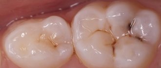
- Fissure. The lesion affects the grooves of the chewing surface.
- Interdental. The development of the disease concerns the surfaces of units in contact with each other. The form proceeds secretly for a long time.
- Cervical. Localization - the area between the crown and gum. Externally, the pathology is visualized as a yellow-brown band at the base of the tooth.
- Circular is the most severe form of flow. The tooth is damaged evenly, completely. The only treatment method is usually surgery.
- Hidden. Located in areas that are inaccessible during a dental examination.
Photo 1. Cervical caries. The process affects the cervical area of the tooth: the neck and the area bordering the gum.
Coming from the depths
- Initial : a grayish-chalky stain on the surface of the enamel.
- Superficial : the enamel is softened, the tooth is sensitive to cold and hot irritants.
- Medium : dentin is involved.
- Deep : in addition to dentin, the pulp is affected, then periodontitis develops.
Due to the sequence
- Primary - defeat for the first time.
- Secondary - pathology occurs after treatment around the filled areas.
- Recurrent - under a filling.
From the point of view of pathological anatomy
- White spot.
- Pigmented spot.
Photo 2. Caries in the white spot stage develops due to the softening of hard dental tissues, the tooth begins to lose its natural shine, which indicates demineralization of the enamel.
Treatment of caries in children
Childhood caries and its treatment still remain an open question for some parents. Even knowing that the child has caries, they are in no hurry to see a doctor, thinking as follows: “Dairy teeth will fall out anyway.” Such judgments are absurd, because infected baby teeth will lead to complications in the growth of permanent teeth or provoke an exacerbation of other diseases in the baby’s body. One way or another, you will have to remove the baby tooth ahead of time. Caries in pediatric dentistry is considered one of the most common diseases, so both parents and specialists should ask questions about how and why to treat caries in children. Dentists believe that treating caries in a baby tooth will preserve it until the molars appear.
If the doctor finds that a child has incipient caries, then most likely, treatment of baby teeth will not be required. The specialist will protect the enamel from further destruction using a painless and effective procedure - applying fluoride varnish or a silver fluoride compound. Then, when the baby gets permanent teeth, the dentist can seal the fissures - the depressions between the elevations of the tooth, which will prevent the occurrence of bacterial plaque that destroys the enamel. If caries progresses, you can no longer do without filling. The doctor will quickly and almost painlessly remove the affected tissue and hermetically seal the tooth. Otherwise, caries can turn into pulpitis in a child, and then into periodontitis.
Medical Internet conferences
Relevance. As is known, today caries is one of the most common diseases in the world (over 95% of people) [1-4]. Diagnosis and prevention of the development of the carious process are still considered important and incompletely studied problems in modern dentistry. It has already been proven that caries is a multi-stage process [5-8]; For a cavity to form, a combination of risk factors and time is required. It often happens that during an appointment with a dentist it is not always possible to diagnose the carious process or the risk of caries. In most cases, the patient, having turned to a doctor for help, already has carious cavities, which leads to the preparation of tooth tissue and subsequent filling. The task of dentists today is to maximize the preservation of their own tooth tissues and prevent the pathological process at an early stage of its development. There are various objective tests for identifying a cariogenic situation (KOSRE, TER test, CRT test), various methods for identifying caries (basic and additional), but when used separately they are uninformative and questionable [9-13]. Diagnosis of caries in a more accessible and faster way at the early stages of its development remains one of the pressing problems in modern dentistry. This article provides a comparative description of various methods for diagnosing caries, taking into account their advantages and disadvantages.
Purpose: to conduct a comparative description of methods for diagnosing caries.
Tasks:
1. Study basic and additional methods for diagnosing caries.
2. Study modern methods for diagnosing caries.
3. Compare standard and modern instrumental methods for diagnosing caries.
Materials and methods. An analysis of relevant literature, scientific articles and dissertations was carried out.
Results and discussion. Diagnostics is an important aspect of clinical medicine, without which it is impossible to make a diagnosis, therefore, the subsequent prescription of treatment and preventive measures is difficult. Primary importance in identifying caries is given to early diagnosis, when the patient does not complain. This is due to the fact that previously identified defects are easier to eliminate, and thus it is possible to prevent the progression of the pathological process. Due to the early manifestation of the carious process, in the absence of diagnosis, an increase in the intensity of dental caries is possible from KPU 2.7 (2004-2006) to KPU 3.5 (2011) [2] in all age groups of the population. It should be assumed that the fundamental solution to this problem is the study of modern approaches to diagnosis and its subsequent implementation to identify the early stages of the carious process [14-16].
The main methods for diagnosing caries include: questioning (disease history, life history), visual examination, probing, percussion, determination of tooth mobility. There are also additional methods, such as vital staining, radiography, selective separation of teeth (access to the proximal surfaces of the lateral teeth), electroodontometric diagnostics, ultrasound, etc. [17-19]. Visual examination is considered the very first clinical method for diagnosing caries used in practice. Until 1920, it involved a combination of visual and tactile examination of teeth using a probe, and clinical diagnosis was based exclusively on this method. But the disadvantages of a visual examination outweigh the advantages: insufficient information content, inability to identify the maximum number of carious cavities. Due to the rapid development of dentistry in general, it would be a mistake to rely only on examination when making a diagnosis, so it is combined with other diagnostic methods (vital staining, radiography).
The vital staining method is used for the differential diagnosis of caries and non-carious lesions. This method is based on the penetration of a dye (1% solution of methylene blue, 0.5% solution of methylene red) into demineralized enamel at the initial stage of the pathological process, when enamel permeability increases due to an increase in the number of pores, thereby the dye is absorbed and the lesion becomes colored in the color of the dye. This method is very convenient, visual and economical, but also has its disadvantages: the inability to assess the depth of the lesion, difficulties in diagnosing caries in hard-to-reach surfaces.
Visual diagnosis remains one of the main methods for diagnosing caries to this day, but more and more specialists are recognizing that basic methods are not enough to identify early carious lesions, especially in hard-to-reach places. Therefore, the solution to this problem was the introduction into clinical practice of instrumental methods for diagnosing caries. The very first and easily accessible instrumental method is radiography, which is quite widely used today. The following radiological methods are most often used in dental practice: intra- and extraoral radiography, survey radiography, long-focus radiography. Interproximal radiography, as one of the reliable methods, refers to intraoral radiography and allows you to obtain an image of the marginal sections of the alveolar processes of the upper and lower jaws without distortion, and to visualize defects in the approximal surfaces of the teeth. Radiography, as a method for diagnosing caries, makes it possible to identify hidden and secondary carious cavities, but has a number of disadvantages: the negative effect of ionizing radiation, the inability to identify initial caries and foci of enamel demineralization, difficulties in determining the depth of damage by the pathological process, and static images. Despite all the shortcomings of x-ray diagnostics, scientists are developing new methods that doctors are already using and will use in the near future, but so far they are rare and remain expensive.
The next method used in the clinical practice of dentists is electroodontodiagnosis (EDD), based on determining the sensitivity threshold of the pulp to electric current. The essence of EDI is to irritate the tooth pulp with electric current and determine the minimum current strength before the first mild pain appears. Thus, it is possible to differentiate the forms of caries and identify its complications. The disadvantages of this method include: the inability to detect initial caries, determine the depth, topography and degree of activity of the carious process, the difficulty of working with the device and interpreting the results. Currently, electroodontodiagnosis is considered an inhumane method for identifying caries in children due to the pain reaction.
The laser fluorescence method using the DIAGNOdent diagnostic device (KaVo, Germany) allows us to identify changes in the structure of dental tissues during the process of demineralization, mainly on the occlusal surfaces of teeth. The laser photodiode of the device emits light waves with a length of 655 nm (red radiation) and a threshold power of 1 mW onto the tooth surface. Organic and inorganic molecules of hard dental tissues absorb light, and the device is reflected in the infrared spectrum. As a result, the device displays values in numbers and notifies with an audio signal. For higher accuracy of readings, it is recommended to clean and dry the tooth before diagnosis. If oral hygiene is unsatisfactory or there is heavy plaque, the device may produce incorrect values. The main advantages of this method are ease of use, absence of harmful ionizing radiation, identification of hidden carious cavities, recognition of fissure caries. Also, with the help of digital and sound identification, you can clearly determine the severity of the disease. However, the device is not intended for diagnosing the contact surfaces of teeth, since in most cases it is not possible to insert the tip of the device into the interdental space. This significantly reduces the scope of application of this device [20-22].
Another option for diagnosing caries is the quantitative light fluorescence method (Quantitative Light-induced Fluorescence, QLF method). The quantitative light-induced fluorescence apparatus is based on a decrease in the ability of hard dental tissues to fluoresce during demineralization. The device is a portable system for intraoral examination with a non-coherent light source and a filter system to replace the laser source. The light-emitting system generates blue light with an intensity of 370 nm, which is transmitted through a liquid-filled light guide. During the examination, the tooth absorbs a blue pulsed stream, thereby healthy teeth glow green, and those affected by caries glow red. The image of the fluorescent tooth is transmitted to the monitor using a video camera through a high-frequency filter. A color image is displayed on the screen showing the condition of the patient's oral cavity. The device is designed for early detection of carious lesions due to the loss of fluorescence in areas of demineralization, determining the location, depth and size of the carious cavity, as well as the severity of the pathological process.
Thus, the introduction into clinical practice of new methods for diagnosing caries will prevent further development of the carious process in the early stages, and will also facilitate treatment using non-invasive techniques without preparation while preserving the tooth’s own tissues.
Table 1.
| Properties | Methods for diagnosing caries | ||
| Visual inspection | Vital coloring | Radiography | |
| 1. Time costs | minimum | minimum | average |
| 2. Information content | insufficient | average | average |
| 3. Cost-effective | + | + | + |
| 4. Complexity of work and interpretation of results | — | — | — |
| 5. Detection of hidden cavities, secondary caries | — | — | + |
| 6. Determination of the location and depth of the lesion | + | — | — |
| 7. Determination of the degree of activity of the lesion | — | — | — |
| 8. Possibility of getting incorrect results | high | high | minimum |
| Properties | Methods for diagnosing caries | ||
| EDI | DIAGNO-dent | QLF method | |
| average | minimum | minimum | |
| 1. Time costs | average | high | high |
| 2. Information content | — | — | — |
| 3. Cost-effective | + | + | + |
| 4. Complexity of work and interpretation of results | + | + | + |
| 5. Detection of hidden cavities, secondary caries | + | + | + |
| 6. Determination of the location and depth of the lesion | — | — | + |
| 7. Determination of the degree of activity of the lesion | — | + | + |
| 8. Possibility of getting incorrect results | minimum | high | minimum |
Conclusions:
1. The main method of detecting caries is a thorough visual examination using a dental probe and mirror. Additional methods include vital staining, radiography, and electroodontic diagnostics (EDD).
2. Modern instrumental diagnostic methods include the laser fluorescence method using the DIAGNOdent diagnostic device (Kavo, Germany), and the quantitative light fluorescence method (QLF method). Hardware diagnostic methods make it possible to identify and evaluate carious lesions at the earliest stages, which is a huge advantage in prescribing timely treatment using non-invasive methods for treating caries.
3. A comparison of basic and additional methods for diagnosing caries showed that there is no ideal method for detecting the carious process with adequate sensitivity and specificity for all tooth surfaces. The most effective is an integrated approach to the clinical situation. A combination of several diagnostic methods is acceptable, the choice of which depends on the tooth surface being assessed. All of the above instrumental diagnostic methods are in addition to a clinical visual examination and are used to clarify the diagnosis.
Features of caries treatment in children
- Before administering anesthesia, the doctor must numb the injection site using special gels or spray. Moreover, the content of the anesthetic component should be minimal. Anesthesia is, as a rule, carried out only in cases where pulp removal is required - that is, with medium and deep caries.
- It is advisable to remove affected tissue using hand tools or a drill, taking frequent breaks.
- As fillings, materials that can be applied “one time” should be used in order to reduce the operation time.
- If caries is complicated and its treatment requires the necessary filling of the roots, then the canals are disinfected without special mechanical treatment and filled with a special paste.
- Treatment of baby teeth in children should take no more than half an hour, otherwise the child will get tired.
- Treatment of multiple, or bottle, caries, as a rule, takes place in several stages and requires the use of sedation.
- Dental treatment under general anesthesia in children is carried out strictly according to indications.
You can get rid of carious lesions at the initial stage using modern methods of caries treatment without a drill. Comfortable, painless treatment is suitable for those children who are afraid of manipulation with a dental bur. The cost of treating caries in baby teeth without a drill is slightly higher than the price of traditional dental treatment for children, but it is worth it. If simple caries gives complications, then you will have to resort to classic drilling. The cost of treatment of complicated caries of primary teeth will depend on the degree of neglect of the disease.
General information
According to medical statistics, 70-80% of children under the age of 10 suffer from caries.
Even children aged 1-2 years are susceptible to the disease, in which areas near the neck of the incisors and canines are affected. Caries quickly spreads throughout the mouth of children. This is due to the characteristics of children's saliva, which does not have antiseptic properties. Bacteria multiply quickly, attacking tooth enamel. Depending on the depth of the cavity defect, uncomplicated and complicated caries are distinguished. The latter is characterized by the development of pulpitis (inflammation of the pulp), periodontitis (inflammation of the tissue between the root and the tooth socket). Depending on the course, dental caries in children can be acute or chronic. The first option is also common in childhood and requires the intervention of a dentist.
Prevention of caries in children
Prevention of childhood caries includes several areas, which boil down to proper nutrition, as well as home and professional hygiene.
Individual oral hygiene
Home hygiene means that as soon as the first tooth hatches, a brush for the baby should appear in a glass next to the parents’ brushes. The same applies to toothpaste. At first, the paste can be applied to gauze or a fingertip to clean your teeth so as not to injure your delicate gums. You need to clean your teeth from all surfaces vertically, then wipe them with a brush soaked in water. Thus, firstly, plaque will be removed, and, secondly, parents will teach the baby to take care of the oral cavity.
Preventing caries with a well-chosen diet
As for nutrition, parental duty is to breastfeed the baby from the very beginning. There is no need to talk about the beneficial properties of breast milk, and the sucking process greatly affects the developing jaw system. Then the baby should be accustomed to fermented milk products. By six months, the child should be fed kefir, and later - cottage cheese and cheese. Parents should remember that the main formation and formation of permanent teeth occurs up to 3 years of age. This means that calcium must always be present in the daily diet.
Professional oral hygiene
This type of prevention involves periodic visits to the dentist and hygienist. The first visit should occur on your first birthday. The specialist will not only give recommendations for care, but also create a diet regimen and examine the mouth. When the first teeth emerge, you and your child will be shown how to brush their teeth properly at an appointment with a hygienist. If there is a problem such as tartar in children, you cannot do without professional hygienic cleaning. It is important for parents to make it a rule to visit the doctor twice a year. And if there are problems with teeth, then more often - once every three months. This is largely due to the fact that various pathologies develop too quickly in childhood. Meanwhile, early diagnosis will lead to quick, painless and relatively inexpensive treatment of caries.
As you can see, in most cases, when caries can attack children's teeth, parents are able to resolutely rebuff it. Of course, there is no escape from genetic predisposition. But it’s not without reason that dental experts say that sweets are the main cause of the development of the disease! So isn’t it better to eliminate the constant source of bacteria and, instead of harmful carbohydrates, offer the child fruits, which are much healthier, after the main meal? Ultimately, your child will thank you in the future for being vigilant and maintaining healthy teeth!
Detailed information and registration by phone: +7 (391) 200-1000.
Make an appointment
Request a call
Prevention measures
Measures to prevent caries in primary teeth include:
- giving up cigarettes, drinking alcohol and other bad habits during pregnancy;
- exclusion of early introduction of artificial complementary feeding (together with mother’s milk, a sufficient amount of nutrients necessary for the full growth of teeth enters the child’s body);
- regular hygiene procedures, which boil down to cleansing the surface enamel layer of teeth from food debris and soft plaque using special children's toothpastes and brushes;
- avoiding the early introduction of sweet foods into the child’s diet, especially carbonated drinks and chewing candies.
In addition, a significant preventive measure is regular (at least once every 6 months) visits to the pediatric dentist and compliance with all recommendations given by the doctor.
Principles of treatment of superficial caries
When superficial caries is detected in children, treatment should be aimed not only at eliminating formations on the tooth enamel, but also at preventing the re-development of the pathology. To prevent complications, comprehensive and immediate therapy is necessary.
When talking about treatment, it should be understood that it is just beginning in the dentist’s office, and it will also need to be continued at home using special means and traditional medicine recipes.
Features of treatment in the pediatric dentist's office
If baby teeth are damaged by caries, then it is quite possible to treat them conservatively without the use of drills, which almost every child is afraid of. Modern dentistry offers more gentle and comfortable methods:
- Children under 3 years of age undergo silvering or deep fluoridation of their teeth. If the enamel has already been damaged by caries, it is cleaned of plaque and treated with a laser. If necessary, cracks in the chewing and frontal teeth are sealed;
- at the age of 3 to 5 years, ozone therapy can be performed to eliminate the first stage of superficial caries, and in case of major complications, treatment is carried out using the method of depophoresis. Its essence lies in the introduction directly into the root canals of a special composition with calcium and copper;
- Children from 5 to 9 years old undergo filling. Glass ionopolymers or colored compomers are used for fillings;
- Children aged 9 to 12 years already have molars and adult technologies can be used to treat them with minimal trauma. But in most cases, therapy of chewing teeth is carried out by sealing fissures.
When superficial caries in children requires complex dental procedures, local anesthesia is mandatory before such procedures are performed. If removal is necessary, such an intervention can be performed under intravenous anesthesia.
Treatment at home
Along with professional treatment of caries by a pediatric dentist, proper oral care at home will also be required to obtain an effective result. For this purpose, special rinsing compositions and toothpastes with anti-caries effects are used.
For children under 4 years of age, dental and oral care products should not contain fluoride. The therapeutic effect is provided by antibacterial active components and calcium ions.
Children over 4 years old are allowed hygiene products containing fluoride, only the concentration of the element must be minimal.
LACALUT kids, PresiDENT Baby, SILPA Putzi, Splat junior are considered good toothpastes for children. You need to brush your teeth with them twice every day. Among the rinses, Drakosha for children, Active Kids, and LACALUT Teens have proven themselves well. When treating caries with such solutions, it is necessary to rinse your mouth after every meal and always before going to bed.
In addition to professional medications, you can also use remedies prepared according to alternative medicine recipes:
- An infusion of chamomile is perfect as a rinse. To do this, pour a glass of boiling water into 1 tbsp. l. dried flowers, leave and cool to room temperature;
- a weakly concentrated solution of sea salt has a positive effect on the condition of teeth. Dissolve 0.5 teaspoons of salt in a glass of water and use for rinsing.
Important! Rinse aids can only be used after the child reaches 1.5-2 years of age, when he can already rinse and spit out liquid on his own, rather than swallow it.
