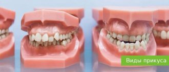
Classification
In orthodontic practice, various assessment criteria are used to determine which dental bite is correct and how to correct existing anomalies. The ideal situation, in which nothing needs to be corrected, is characterized by the following indications:
- Semi-elliptical shape of the upper and parabolic shape of the lower arc.
- Small - no more than a third - overlap of the vestibular surface of the mandibular elements.
- Contact of antagonists during closure and absence of obvious gaps.
Compliance on all points is quite rare, but in most cases the deviations are minimal and do not require medical intervention. Often the cause of imbalance is not congenital defects, but bad habits, some of which are formed at an early age.
Types of correct bite
In accordance with the generally accepted classification, there are four main types of occlusal relationships, characterized by the absence of problems and not having a negative effect on the jaw region.
Orthognathic
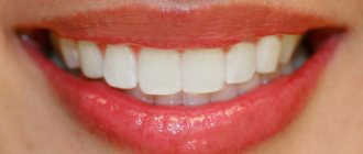
The optimal, from an aesthetic point of view, is a condition in which all elements of the dentition have an even shape, are located in the correct manner, without gaps or deviations from the midline.
Straight
A common phenomenon, the main feature of which is the lack of overlap - the cutting edges of the incisors meet along a line, which causes a specific type of smile, but is not considered as a pathology.
Biprognathic
Another type of even dentition without threes and diastemas, the distinctive feature of which is a slight deviation of the vertical growth vector of the units. With this development, the crowns are slightly tilted forward.
Progenic
In a normal state it is not accompanied by abnormal manifestations. The identifying feature is a slight frontal protrusion of the lower units.
Types of normal occlusion
According to statistics, no more than 30% of the population have a normal bite. There are several types of teeth arrangement that meet all the signs of a correct bite:
- Orthognathic. The teeth of the upper jaw overlap the lower teeth by a third of the crown size. When the jaws close, close contact occurs between the teeth.
- Straight. The cutting edges of the upper and lower teeth are in close contact. Correct occlusion of molars and premolars is mandatory.
- Biprognathic. The upper and lower incisors have a slight inclination towards the vestibule of the mouth. There is contact between the incisors, and the upper canines slightly overlap the lower ones.
- Progenic. The teeth of the lower jaw are pushed forward relative to the upper ones. The closure is maintained.
With any type of normal positioning of teeth, bite correction is not carried out, since the load on the teeth is carried out evenly. Food is chewed thoroughly, the temporomandibular joint is not overloaded.
Which teeth bite is not considered correct?
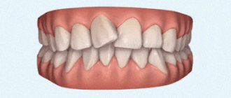
The presence of deviations from the norm is at least the basis for a dental diagnosis, based on the results of which a decision is made on the need for treatment. The best option is orthodontic correction in childhood, since the dentofacial apparatus, which is in the stage of formation, is more easily amenable to the targeted influence of a special machine and takes on the desired structure without surgical intervention. The most commonly diagnosed abnormal forms have certain signs that allow us to understand whether a person’s bite is correct or not, what is going wrong, and how this deviation can affect the further development of the jaw:
- Distal - diagnosed with excessive development of the upper jaw, noticeably protruding forward and distorting facial features.
- Mesial is a reverse pathological condition in which the mandibular row protrudes, almost completely covering the incisors and canines.
- Cross - characterized by the presence of free areas between the teeth, as well as a “scissor” intersection of elements in a random order.
- Deep - significant, more than 30%, overlap of the lower section.
- Open - contact during closure is observed only between individual chewing units, while in the incisal region there are pronounced gaps, creating the impression of a constantly slightly open mouth.
Any pathological manifestation requires timely medical intervention. Not all patients know what a correct bite in a person is and what it is needed for - but this indicator affects the quality of respiratory and speech function, ensures the conditions for normal food processing and the functioning of the gastrointestinal tract. By contacting Dentika dentistry, you can undergo a comprehensive diagnosis, receive recommendations from qualified specialists and, if necessary, resort to orthodontic correction services.
Causes of pathological bite
The formation of occlusion is a long and complex process that begins at the stage of intrauterine development, continues in early childhood and ends after the end of puberty. Pathological occlusion is the result of long-term influence of a number of unfavorable external or internal factors at different periods of a person’s life:
- Genetic factor. A genetically determined anomaly is formed when there is a congenital discrepancy in the size of the teeth and dentofacial bones (crowding of teeth or the presence of spaces between teeth) or a hereditarily determined abnormal shape and size of the lower and upper jaws.
- The influence of negative factors on the body of a pregnant woman. The formation of the dental apparatus in the fetus can be influenced by bad habits, poor nutrition and diseases suffered by a woman during pregnancy.
- Birth injury. During childbirth, the baby may receive injuries leading to displacement of the temporomandibular joint.
- Negative factors affecting the baby’s body in the first years of life. The reason for the formation of a distal bite may be the incorrect selection of nipples or the late introduction of solid foods into the diet.
- Bad habits and behavioral factors in early childhood. The habit of sucking a pacifier or finger, or sleeping with the head thrown back can cause a child to develop a deep bite.
- Factors influencing the formation of anomalies in later life. These include carious lesions of the teeth, ENT pathologies, deviated nasal septum, and diseases of the skeletal system.
Treatment of abnormal bite should begin as early as possible. Early diagnosis and therapy make it possible to restore the normal functioning of the dentofacial apparatus more effectively. Doctors at the Consilium-Dent use specially developed diagnostic tests and tables. They are used both for direct examination of the oral cavity and for diagnosis using dental casts.
The need for a particular therapeutic measure is determined by a specialist based on the diagnostic results on an individual basis. You cannot resort to self-medication in this situation. This can lead to activation of the pathological process.
Treatment for missing teeth
The specificity of the anatomical structure of the jaw apparatus is that even complete edentia does not guarantee the formation of correct occlusion after implantation. The result of prosthetics is largely determined by the degree of development of defects and anomalies that formed even before the loss or removal of elements. If the row ratio was within acceptable limits, it is enough for the doctor to take into account the necessary adjustments when developing a replacement structure, but the presence of problems with bone tissue will require the use of more complex restoration techniques.
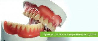
How to check and find out if the bite is correct when making a jaw prosthesis without teeth? For this purpose, special wax rollers are used, placed in the oral cavity. The measuring instruments are a ruler arc and an intraoral plate. After collecting the necessary readings, first a plaster prototype is created, and then the final structure.
Numbering of teeth in dentistry
Numbering of teeth in dentistry
In world dentistry, several alternative tooth numbering options are used. In European countries, which includes the Russian Federation, the European International two-digit numbering system Viola is widely used. In America, the alphanumeric system is used. There is also the Haderup technique, in which the elements of the upper and lower row are designated by the symbol “+” or “-”, the Zsigmondy-Palmer scheme and the universal system, in which each element is assigned a serial number, and the numbering goes from right to left, starting with the right upper molar.
Practical meaning of numbering
From a practical point of view, tooth numbering in dentistry is very important. Firstly, it is necessary to fill out medical documentation, which is maintained by all dentists. Secondly, without it it is difficult to imagine coordinated interaction between doctors with different specializations (dental therapists, prosthetists, orthodontists, surgeons). How to inform a colleague about what elements of the dentition have been treated, especially when the patient receives treatment in different clinics? With the help of a single scheme this is quite simple.
A separate area of application of tooth numbering in dentistry is diagnostics. Numbering is used when describing a panoramic image or teleradiography. It is even more important when performing computed tomography, because a 3D image does not always allow one to understand which element of the dentition is depicted.
Finally, numbering simplifies communication between patient and specialist. Of course, the patient can explain and even show the doctor which tooth is bothering him. However, it is much easier to do this using a universal nomenclature or the correct names of teeth, with which we will begin a more detailed study of this issue.
What are the teeth called?
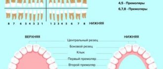
Each tooth has a unique name. The four front ones - two left and two right - are usually called incisors. Immediately behind them are the fangs. Behind the fangs begin the elements of the chewing group - premolars. The lateral elements are usually called molars.
Incisors are usually divided into medial (central) and lateral (lateral). Accordingly, each person has an upper left medial incisor and a left lateral, a lower central left and a lateral. The same exact elements are located on the right.
With fangs everything is much simpler. There are top left and right, bottom left and right. There are four fangs in total in the oral cavity, regardless of whether they are baby teeth of a child or molars of an adult.
There are two premolars in each half of the dentition. They are designated by a serial number, starting from the center line - the first left premolar from above, the second from the left, and so on.
There are the most molars - twelve in total. When designating them, the same principle is applied as when numbering premolars. An element of the dentition is named by a serial number indicating its location - in the upper or lower row, on the left or on the right.
Milk teeth have a significant difference. The child has no premolars, only two molars on each side. Accordingly, there are eight molars in total, and they should be named the same as those of an adult.
Viola system: European two-digit circuit
As we can see, even without any numbering, the patient can explain to the dentist exactly which tooth hurts. But it won’t be possible to describe a panoramic photograph of the jaws or fill out a medical card - the description will be too cumbersome. The orthodontist will encounter even more problems when installing braces. Each lock is designed for a strictly specific tooth, and it must be clearly indicated for which one.
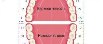
To simplify the work of dentists and, more importantly, eliminate the possibility of error, the two-digit Viola numbering system was developed in 1971. The dental system is divided into four segments or quadrants. The right quadrant of the top row received index 1, the top left quadrant received index 2, the bottom left quadrant received index 3, and the bottom right quadrant received index 4. Thus, the first digit in the designation of a particular tooth refers to the segment in which it is located.
- 1 - right side of the top row.
- 2 - left side of the top row.
- 3 - left side of the lower jaw.
- 4 - right side of the lower jaw.
The second number indicates the location of the element in the dentition. Numbering begins with one and proceeds from the central incisors to the third molars. Accordingly, the lateral incisor is designated by the number 2, the first premolar by the number 4. The wisdom tooth receives the number 8.
By combining indices (designation of a segment) and serial numbers (indicating a specific tooth), we obtain a universal system that eliminates the possibility of error. For example, 32 is the lateral incisor of the left half of the lower jaw. 18 - third molar of the right half of the upper row. Obviously, such a nomenclature greatly simplifies both patient management in a dental clinic (stages of diagnosis, treatment) and communication between specialists.
Practical examples
Try giving a name to the maxillary canine on the left. The correct answer is 23. The index “2” is assigned to the element of the left half of the upper jaw. The number three is the number of the element in a given segment of the dentition. Fangs come third.
Another task. Which element is numbered 45? Index 4 unmistakably indicates the right half of the lower jaw. The second digit is the serial number. Fifth are the second premolars. So, 45 is the second right premolar of the lower row.
Viola system and baby teeth
The universalism of the international Viola system becomes obvious when describing teeth in children. For primary teeth, other indices are used:
- 5 - right half from above.
- 6 - left side of the top row.
- 7 - left half of the bottom row.
- 8 - right half of the bottom row.
The second digit indicates the element's serial number. Accordingly, 55 is the second primary molar on the upper jaw on the right. 71 - central incisor of the lower jaw on the left.
It happens that a child has already erupted some permanent teeth, but still has milk teeth. In such a situation, numbering according to the Viola scheme is very convenient. The dentist will write down 31 and 72 in the medical record. What does this mean?
This means that the central incisor of the left half of the lower row (31) has already been replaced by a permanent one, while its neighbor, the lower left lateral incisor (72), still remains one of the baby teeth.
The Viola numbering system was adopted by WHO and is recognized as the most universal, and therefore we paid maximum attention to it. We will not analyze other schemes in such detail, since they are used much less frequently in Moscow clinics.
Other numbering systems
Universal system. You can not divide the dental system into squares, but simply designate the elements by their serial number. The count starts from the upper right third molar and goes counterclockwise in a circle. Accordingly, 8 is the upper central incisor on the right, 10 is the left canine on top, 17 is the wisdom tooth in the bottom row on the left, and 25 is the medial incisor of the lower jaw on the right.
For baby teeth, the universal system uses not numbers, but letters of the Latin alphabet from A to T, which is not very convenient. The letters from A to E indicate the upper milk teeth on the right, from F to J - the left upper milk teeth, from K to O - the lower left milk teeth, from P to Q - the right milk teeth from below.
Haderup system. At first glance, this is a simple and convenient numbering system, but due to the constant use of the same numbers, only with different indices, when using it, the likelihood of error increases. According to the Haderup system, teeth are designated as follows:
- numbers from 1 to 8, where 1 is the central incisor, and 8 is the third molar;
- indexes + or -, where “+” denotes the teeth of the upper jaw, “-” denotes the teeth of the lower jaw;
- for baby teeth, 0 (zero) is placed before the serial number;
- when designating the teeth of the right half, the index + or - is placed to the right of the number;
- when designating the teeth of the left half, the index + or - is placed to the left of the number;
Examples of numbering according to the Haderup system:
- 8+: right maxillary third molar;
- -1: left mandibular central incisor;
- 02-: lateral primary incisor of the lower jaw on the right.
Alphanumeric system. The system was proposed by US dentists. In this numbering scheme, each type of teeth receives its own capital letter of the Latin alphabet, and the serial number of the element in a given group of teeth is indicated by a digital code. Baby teeth are numbered in exactly the same way, only small letters are used instead of capital ones:
- I (i) - incisors.
- C (c) - fangs.
- P - premolars (not in children).
- M (m) - molars.
Examples: I2 - lateral incisor. M3 - third molar. The first primary molar is numbered m1.
The disadvantage of the alphanumeric system is that it does not indicate in which jaw the element of interest to the doctor is located; it does not take into account whether it is on the right or left.
There are other systems for numbering teeth in dentistry, but they are considered imperfect and are practically not used today. In order not to confuse the reader, we will not dwell on them.
If you have questions regarding the timing of the eruption of primary and permanent teeth in children, oral hygiene, correction of malocclusions, diagnosis or treatment of dental diseases, ask them to a dentist at the Galaktika clinic (Moscow) during an individual consultation.
Treatment of malocclusions
Before prescribing treatment procedures, the orthodontist assesses the condition of the oral cavity, carefully examines the depth of the problem and the presence of concomitant pathologies, taking into account the patient’s age. Treatment of dental anomalies is carried out using conservative and radical methods:
- Braces are the most popular method of bite correction. This orthodontic method has the widest range of applications and is suitable for solving any problem of malocclusion. Treatment with this method is long and includes preparatory, main and retention (recovery) periods.
- Aligners. Removable structures are used to treat the initial stages of deviation. With their help, a gentle correction of the dentoalveolar apparatus is carried out, changing one set of mouthguards to another.
- Application of veneers, onlays, crowns. Weak areas of the dentition that are susceptible to caries are protected by using orthopedic structures. Chipped areas of enamel are restored using ceramic onlays and veneers.
- Surgical intervention is performed to treat late stages of dental anomalies (mainly of a hereditary nature). During the operation, the position of the jaws is changed, their size is reduced or increased.
Laser therapy is performed as an auxiliary treatment to help speed up regeneration and reduce the risk of complications.
Histological structure of teeth
All the most interesting things about human teeth can be learned using histology. Histology is a science that studies the morphological structure of organs and systems at the tissue level. Human teeth and roots consist of hard (97%) and soft (3%) tissues. Hard tissues are represented by dentin and enamel, and soft tissues are represented by vessels and nerves of the pulp, located in the so-called pulp chamber and root canals, inside the coronal and root parts of the tooth. The basis of the tooth is dentin.
The coronal part of the crown is covered with enamel on top of the dentin, and the root part, located in the bone, is covered with cement on top. The border where the root cement approaches the enamel of the coronal part is located in the area of the tooth neck. The roots of the tooth are attached to the bone tissue of the alveolar process of the jaw with the help of periodontal fibers, which is called the ligamentous apparatus of the tooth. With its help, the tooth can withstand chewing loads. The abundance of nerve fibers in the pulp is the cause of intense toothache with deep caries and pulpitis.
Common types of malocclusions
The most common anomalies of the dental system include deep bite. A severe degree of pathology is characterized by a situation where the upper row of teeth completely overlaps the lower row. When the jaws are closed, the upper incisors can injure the mucous membrane of the lower gums. Therefore, this type of anomaly is called a traumatic bite.
A deep bite is accompanied by a violation of facial aesthetics, impaired diction, poor quality of chewing food, and increased abrasion of the enamel.
There are two degrees of development of pathology. They are caused by various provoking factors and are characterized by different forms of manifestation:
- Primary deep bite is often due to genetic reasons and is determined by the special structure of the facial bones. It manifests itself as pathological changes in the area of the dentition, alveolar process and basal part of the jaws. The movement of the jaws is blocked, and the patient experiences significant discomfort when chewing.
- Secondary deep bite is acquired in nature and is a consequence of a disease or injury. As a result of a decrease in the interalveolar distance and the lower third of the face, a deep traumatic closure of the incisors develops.
Another type of pathology classification is carried out depending on the nature of the interaction of the lateral teeth.
- A deep distal bite is characterized by the overlap of the lower row of teeth with the upper ones by more than one third of the length of the crown with a violation of the relationship of the lateral teeth. Characteristic disorders include difficulty biting and chewing, breathing problems, and pain in the temporomandibular joint.
- Deep neutral bite: when the overlap of the lower front teeth with the upper ones is disturbed, while the position of the lateral teeth is not disturbed.
If you experience even minor discomfort when chewing, you should immediately contact a specialist and follow his recommendations.
The presence of a malocclusion or a predisposition to it is determined by an orthodontist after an initial examination using special tests. Self-diagnosis does not provide an objective picture of the condition of the dentofacial apparatus, since different pathologies may have the same symptoms. Treatment in this case will be radically different.
If there are no problems with your bite, you should visit the orthodontist for a preventive examination approximately once every six months. The doctor will determine whether there is a tendency to a particular anomaly even before the problem is noticeable.
Dental formula
Human teeth are arranged symmetrically in the form of two dentitions - the upper dentition and the lower dentition. There are eight teeth on each side of the dentition from the median plane.
A person has 4 types of teeth:
