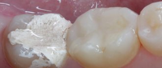
1 - superficial caries; 2- medium caries; 3 - deep caries
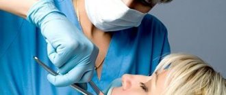
Of course, treatment of caries in modern conditions is always carried out under local anesthesia, so today there is no need to talk about the pain of this procedure at all.
Removal of carious tissue during dental treatment is carried out in a variety of ways.
The most common of them is the preparation of a carious cavity using a drill. This method still remains universal and widely used, despite the fact that it is far from the most favorite for patients in dental clinics.
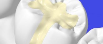
It must be said that along with traditional filling of carious cavities, the Best Dentistry clinic uses sound filling. We use a sonic tip (Germany) for this.
The essence of the method is that a special filling material becomes liquid under the influence of a sound wave and is supplied by a tip under pressure, which eliminates the formation of pores in the filling. When the sound wave is turned off, the material transforms into a thicker and more plastic state, which allows the doctor to model the future composite filling in the most convenient consistency.
Therapy room
Dental treatment can also be carried out using the water-air-abrasive method.
In this case, the removal of carious tissue occurs through a mixture of water, air, and a special abrasive powder. This spray gently removes softened tissue without damaging healthy tooth tissue. Treatment of dental caries using air-abrasive technology is significantly less painful than traditional preparation. Therefore, local anesthesia is usually not required.
In addition to the above, the so-called “chemical” method of treating dental caries using the drug “Karisolv” is used. First, the cavity is treated with Carisolv liquid, and then the demineralized tissue is removed with a hand instrument without a drill. At the last stage, the teeth are filled with a composite material, and dental treatment is considered complete.
Unfortunately, unlike preparing a carious cavity with a drill, the last two methods are applicable only at the initial stages of caries development.
Stages of caries
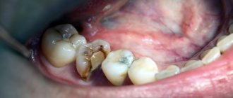
- Initial (superficial, in the stain stage) - a white or brown round-shaped spot forms on the enamel. There is no pain or discomfort. The defect can only be noticed visually. A light color indicates an acute process, and a dark color indicates a chronic one.
- Medium (dentin damage) - the infection eats away the enamel, spreads to the dentin, and forms a cavity in it. Slight pain occurs when biting or applying heat to the tooth. The edges of the cavity are sharp and periodically crumble.
- Deep - the infection reaches the pulp, the tooth begins to disturb constantly, especially at night. The pain is intensified by temperature irritants and sweets. The attacks are strong, but short (last 5-10 seconds).
It is impossible to accurately determine the degree of caries by eye. The degree of damage can be diagnosed only by drilling the tooth. Often, externally, the lesion looks medium, but turns out to be deep, so only the estimated price of therapy is discussed with the doctor in advance.
Contraindications
- Acute stage of ARVI, acute respiratory infections, herpetic rashes
- I, III trimester of pregnancy
- Acute stage of periodontal disease
- Bruxism (night grinding of teeth)
- Seizure attacks
Indications
- Darkening of tooth cusps, cervical areas
- Chipping of a piece of tooth enamel
- Dark gap along the border of the filling
- “Dots”, “spots” on teeth
- Pain from sweet, sour, cold
Indications and contraindications for dental treatment with the Icon system
We have to admit that although the technique is a small miracle in the world of dentistry, since it allows you to treat teeth without a drill, it is not suitable in all cases.
Indications for treatment with Icon:
- fear of dental procedures (dentophobia), anesthesia;
- allergy to anesthesia drugs;
- violations of the integrity of tooth enamel after orthodontic treatment;
- caries that affects the enamel of both the upper and deep layers, as well as when the upper third of the dentin is injured. However, some dentists are skeptical about the treatment of caries with affected dentin with Icon;
- the presence of carious areas in the interdental space and other hard-to-reach places.
Contraindications to treatment:
- individual intolerance to the components of the drug;
- children under 3 years of age, due to the possibility of damaging very fragile children's enamel;
- the product will not help with enamel diseases;
- in case of deep damage to dentin.
Diagnostics
In our clinic, caries diagnosis begins with an examination. The dentist carefully examines each tooth from the outside and inside, lightly tapping it (percussion). If a source of infection is detected, thermometry is performed - exposing the tooth to cold to assess the reaction. Electroodontometry helps determine the degree of infection development - the impact of a microcurrent on the tooth, to which the inflamed pulp will react. Patients are prescribed x-rays to assess the condition of the root tissues and detect hidden cavities.
Technologies and materials
To diagnose and treat caries, the PROFI-Dent clinic uses a wide range of advanced methods and materials:
- digital pantomograph and radiovisiograph;
- safe, effective anesthetics of the latest generation;
- advanced dental units;
- modern surgical equipment (physiodispenser, ultrasound and laser devices);
- durable filling materials with high abrasion resistance (light-curing);
- inlays, veneers, etc.;
- modern calcium- and fluorine-containing preparations for remineralizing therapy;
- the latest restoration techniques.
The use of advanced diagnostic equipment and the latest materials ensures impeccable results. Our dentistry provides turnkey caries treatment, which includes not only the elimination of inflammation and pain, but also the complete aesthetic restoration of the tooth’s shape using light-transmitting materials.
Stages of traditional caries treatment (using a drill)
- Anesthesia completely removes pain. At DHC, local anesthesia is performed with new generation drugs based on Articaine. The injections are completely safe, hypoallergenic, and have no restrictions or side effects.
- Tooth preparation - excision of carious tissues with a drill. It is necessary to remove all affected tissue, otherwise the infection will recur.
- Creation of a cavity with burs of different sizes with cooling so as not to overheat the pulp and dentin. Inside, the cavity is treated with etching gel to improve adhesion to the filling.
- Installation of a gasket - made on the basis of calcium hydroxide, normalizes blood circulation in the pulp, accelerates dentin regeneration. Spacers are needed when the cavity wall is located next to the pulp.
- Filling with photopolymer materials. Such fillings are durable, similar in color to enamel, and can withstand any load.
- Grinding - adjusting the filling height so that it does not interfere with the closure of the teeth and does not disturb the bite.
Minimally invasive methods for treating caries
Early diagnosis, as well as the availability of modern materials and treatment methods, allow dentists today to effectively combat caries disease without resorting to excessive intervention and removing a significant amount of tissue.
The principle of minimally invasive intervention was first proposed in the 90s, and in 2002 it was officially recommended by the International Dental Federation for use in dental practice. Today it is actively used in the work of a modern dentist.
This principle consists of early diagnosis of caries, constant monitoring of risk factors for the development of caries disease, minimal tissue removal, as well as the use of modern filling materials.
Early diagnosis of caries disease plays a significant role. After all, it is the identification of the disease in the early stages of its development that makes it possible to prevent its development, and therefore the removal of a significant amount of tooth tissue. Diagnostics consists of both classical visual assessment and the use of modern diagnostic methods such as laser fluorescence, electrometric method, etc.
Surgical minimal intervention involves modification of classical preparation methods, the use of special instruments, and modern alternative preparation techniques. These include the air abrasive method, the use of a laser, the ultrasonic method, and chemical methods of tooth preparation.
Based on the depth and volume of the lesion, non-invasive and invasive fissure sealing, preventive filling, atraumatic restorative treatment (hereinafter referred to as the ART technique), as well as other techniques that minimize intervention, may be applicable.
So, in the presence of complex anatomy of fissures, as well as their significant depth, high-quality cleaning of the chewing surface becomes difficult for the patient, which becomes the main factor in the development of caries in them. To prevent the development of caries, a non-invasive fissure sealing technique is used, in which the fissure, after preliminary cleaning, is filled with sealant. Initial lesions involving fissures on the chewing surfaces of teeth can be eliminated by the use of invasive sealant, which involves minimal removal of enamel and then filling the exposed fissures with sealant. If the carious lesion spreads to the dentin, preventive filling is required, here we are talking about removing the affected tissue using small burs, as well as carrying out invasive sealing in the remaining fissures bordering the lesion.
If tissue damage is significant, more significant intervention using the ART technique is required. This technique consists of mechanical removal of the affected tissue with hand instruments, followed by filling the cavity with glass ionomer material. Today, this technique is widely used to treat children, people with severe general somatic pathology that does not allow resorting to more radical methods of treatment, and to treat patients who are afraid of the dentist. Also, the ART technique is applicable in conditions where it is impossible to use machine preparation, which is its undoubted advantage.
In addition, there are methods of tunnel preparation, bate-cave preparation, slot preparation, etc. A little about them. If a small carious lesion is located on the contact surface, and the marginal ridge is intact, the tunnel preparation technique is applicable, in which access to the affected tissue is carried out from the chewing surface while preserving the marginal ridge. Bate-cave - preparation is applicable in the presence of a cavity on the chewing surface, in which the lesion extends under the tubercles. The technique involves creating an entrance in the center of the defect, and then removing dentin under the intact enamel using circular movements. Slot preparation is used to remove carious lesions on the contact surface of teeth. With slot preparation, the marginal ridge is also preserved, and access is made from the lingual and buccal surfaces. It should be noted that these techniques are applicable only with good diagnosis and good manual skills of the dentist.
In conclusion, I would like to say that minimally invasive methods help to preserve healthy tooth tissue as much as possible, which allows you to prolong the life of the tooth after treatment, minimize the patient’s discomfort, and therefore make a trip to the dentist more enjoyable.
Intern at the 7th City Dental Clinic
Antanovich Olga Nikolaevna
Non-invasive methods for caries treatment (without drilling)
- Ozone therapy helps in the initial stages. Carious tissues are not removed mechanically; they are only treated for disinfection with ozone, which destroys 99.9% of bacteria. The procedure completely stops the development of the infection.
- ICON - infiltration of infection at the spot stage. The drug penetrates damaged enamel and makes it durable. The tooth surface becomes resistant to bacteria and organic acids.
- Laser therapy is similar to traditional therapy and is effective at all stages. It differs in that damaged tissue is not drilled out, but evaporated by a laser.
- Sandblasting (air abrasive) treatment is indicated for the treatment of initial caries. The sandblasting machine has the same principle of operation as a drill, but without vibration and contact effects on the enamel.
Air-abrasive method of preparation
Essentially, this is sandblasting the hard tissues of a diseased tooth. Through a special tip, an aerosol containing water and an abrasive substance is applied to the surface of the tooth. This method is used before sealing fissures or to remove pigmented areas of tooth enamel.
The air abrasive method can be used to prepare small carious cavities. The advantages of this method are minimal tissue excision and the creation of a rough surface that does not require additional acid etching.
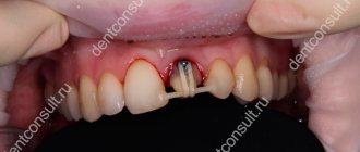
Complications after treatment
If the dentist overheats the dentin or injures the pulp during preparation, the tooth will begin to ache after the anesthesia wears off. We'll have to re-open it and depulp it. A lump may appear on the gum after unsuccessful canal sealing if the material penetrates beyond the root. Then you need to re-open the canal, treat the inflammation, then fill it again.
If the filling is in the way, it means that the dentist did not sharpen or polish it properly. This can be easily fixed. The appearance of a reaction to thermal changes indicates a violation of the tightness of the seal. All these complications often arise due to the fault of the dentist. To prevent them, you should carefully choose a clinic and trust only doctors with extensive experience.