MOUTH, ORAL CAVITY
[
mouth, oral cavity
;
os (oris)
(PNA), os (JNA, BNA),
cavitas oris
(PNA)] - the initial section of the digestive tract, consisting of the oral opening and the oral cavity. The oral opening is limited by the upper and lower lips (see). When the lips are closed, the mouth opening has the shape of a mouth slit (rima oris); when the lips are open, it has a rounded shape. The size of the oral fissure varies and averages 6-8 cm in adults. In men, the oral fissure is usually larger than in women.
In the oral cavity, mechanical grinding of food occurs and its chemical processing begins, preparing the food for further digestion into the gastrointestinal tract. tract. In addition, the oral cavity takes part in breathing, as well as in the formation of speech and singing sounds.
Embryology
During embryogenesis, the formation of the oral cavity is closely related to the development of the face (see). At the head end of the embryo, an invagination of the ectoderm is formed, which grows towards the blind end of the foregut. The so-called the oral fossa, or bay, which is the rudiment of the primary oral cavity, as well as the nasal cavity. During the 6-8th week. During embryonic development, the primary oral cavity is divided into the final oral cavity and the nasal cavity, which is associated with the formation of the hard and soft palate. The formation of the vestibule of the mouth is closely related to the development of the lips and cheeks. At approximately the 7th week of embryonic development, epithelium grows along the upper and lower edges of the primary oral fissure, followed by its immersion into the underlying mesenchyme in the form of an arcuate plate. Along the course of the plate, a gap soon appears; the edges separate the rudiments of the upper and lower jaws from the rudiments of the lips. Initially, the oral opening of the embryo is very wide and reaches the rudiments of the outer ear. Its reduction in size occurs due to the fusion of the edges of the oral fissure and the formation of the cheeks.
Normal appearance, structure and functions of the palate
The palate is the top of the oral cavity in the form of a dome or vault, separating it from the nasal passages. The sky is divided into two parts:
- hard - at the base there is a bone plate covered with a mucous membrane;
- soft - these are muscles covered with a mucous membrane that raise the palatine during swallowing and lower during breathing.
Sky functions:
- respiratory protection;
- participation in the chewing process;
- assistance in the formation of speech sounds.
Normally, both parts of the palate are a soft pink hue. In diseases of the nasopharynx, redness of the mucous membrane occurs. A purple or bluish tint may appear. The acquisition of a yellow color by the palate in a child or adult indicates the presence of pathology.
Anatomy and histology
The oral cavity is limited in front and on the sides by the lips and cheeks; its upper wall is the hard and soft palate (see), and the lower wall is the floor of the oral cavity. The basis of the floor of the mouth is the diaphragm of the mouth, the edges of which consist of the paired mylohyoid muscle (m. mylohyoideus). Above it are the geniohyoid muscles (mm. geniohyoidei), as well as the muscles of the tongue (see). At the back, the oral cavity is connected to the pharyngeal cavity through the pharynx (see).
Rice. 1. Median sagittal section of the head at the level of the oral cavity and pharynx. Rice. 2. Oral cavity, front view (the corners of the mouth are cropped). Rice. 3. Frontal cut of the head at the level of the molars, 1 - hard palate; 2 - teeth; 3 - upper lip; 4 - oral fissure; 5 - lower lip; 6 - vestibule of the mouth; 7 - lower jaw; 8 - sublingual gland; 9 - genioglossus muscle; 10 - geniohyoid muscle; 11 - mylohyoid muscle; 12 - hyoid bone; 13 - pharynx; 14 - tongue; 15—soft palate; 16—the oral cavity itself; 17—frenulum of the upper lip; 18— gums; 19— palatoglossal arch; 20—palatine tonsil; 21—uvula; 22—frenulum of the lower lip; 23—velopharyngeal arch; 24 - transverse palatal folds; 25 - anterior belly of the digastric muscle; 26 - buccal muscle; 27 - fatty body of the cheek.
Anatomically, the oral cavity is divided into the anterior section, or vestibule of the mouth (vestibulum oris), and the posterior section, or the oral cavity itself (cavitas oris propria). The vestibule of the mouth looks like a gap enclosed between the lips and cheeks (in front and outside) and the teeth and gums (back and inside). With the help of interdental spaces and retrodental spaces, the vestibule of the mouth communicates with the oral cavity itself (color. Fig. 1 - 3).
The oral cavity itself is separated from the vestibule of the mouth by teeth and gums (see). When the teeth are closed, it looks like a gap; when the mouth is opened, the oral cavity takes on an irregular ovoid shape. There are individual and age differences in the shape and size of the oral cavity: in brachycephals it is wider and shorter than in dolichocephals. In newborns and children up to 3 months, the oral cavity is very small, short and low. In the oral cavity there are teeth (see), tongue (see), the excretory ducts of the large and small salivary glands open into it (see).
Both the vestibule of the mouth and the oral cavity itself are lined with a mucous membrane that is resistant to various mechanical, chemical and thermal irritants, has high regenerative capacity and is relatively resistant to infection.
Rice. 10-15. Microscopic specimens of the oral mucosa are normal. Rice. 10. Buccal mucosa: glycogen (indicated by an arrow) in the superficial layers of non-keratinizing stratified squamous epithelium, detected using the PAS reaction, cells of the basal layer do not contain glycogen; x 200. Fig. 11. Non-keratinizing stratified squamous epithelium of the buccal mucosa; coloring with azan; x 400. Fig. 12. The mucous membrane of the hard palate with underlying adipose tissue, covered with keratinizing stratified squamous epithelium: the stratum corneum is intensely red in color; coloring with azan; X 140. Fig. 13. Keratinizing stratified squamous epithelium of the mucous membrane of the hard palate: the stratum corneum is intensely red; coloring with azan; x 400. Fig. 14. Gingival mucosa, covered with keratinizing stratified squamous epithelium (the stratum corneum is indicated by an arrow); hematoxylin-eosin staining; x 200. Fig. 15. The mucous membrane of the red border (transitional section) of the lip, covered with keratinizing multilayered squamous epithelium: the stratum corneum is purple; aldehyde-fuchsin staining; x 140.
Throughout its entire length, the oral mucosa is covered with stratified squamous epithelium. The epithelium covering the mucous membrane of different parts of the oral cavity differs. In the area of the cheeks, lips, soft palate, and floor of the mouth, the epithelium under normal conditions does not keratinize. A characteristic feature of the non-keratinizing epithelium of the human oral cavity is its ability to accumulate large amounts of glycogen in the cytoplasm (color Fig. 10 and 11). In the area of the hard palate and gums, the epithelium shows a pronounced tendency to become keratinized. In these areas, on top of the layer of spinous cells there is a granular layer consisting of elongated cells, which contain keratohyalin grains in their cytoplasm. Above, the granular layer passes into the stratum corneum, consisting of completely keratinized and nucleated cells. In the keratinizing epithelium, glycogen is usually absent (color fig. 12-15).
The epithelium of the oral mucosa has a high level of activity of enzyme systems, including enzymes of the tricarboxylic acid cycle (see Tricarboxylic acid cycle), glycosyltransferases.
The lamina propria of the oral mucosa, on which lies a layer of epithelium, forms numerous projections, or papillae, protruding into the layer of epithelium. The distribution of cellular elements (fibroblasts, mast cells, plasma cells, segmented leukocytes) in different parts of the oral mucosa is uneven: the lamina propria of the mucous membrane of the cheeks and lips is richest in cells. Fibrous structures are represented by bundles of interwoven collagen fibers, between which are located argentophilic and elastic fibers. The largest number of elastic fibers is observed in the lamina propria of the mucous membrane of the cheek and palate.
The lamina propria of the oral mucosa, without a sharp boundary, passes into the submucosal layer (submucosa, T.), which is especially well developed at the bottom of the oral cavity. The submucosal layer contains numerous small salivary glands. The muscular plate of the mucous membrane characteristic of the digestive tract is absent here. In the gum area, in the lateral parts of the hard palate and in the area of the palate suture, the submucosal layer is completely absent. In these areas, the mucous membrane is tightly connected to the periosteum of the corresponding bones.
Blood supply, lymphatic drainage and innervation of the walls of the oral cavity are closely related to the vascular and nervous systems of its constituent formations (see Throat, Teeth, Palate, Jaws, Tongue).
Anatomy
Rice.
1. Lips are normal. The shape and size of the lips depend on the individual characteristics of the orbicularis oris muscle, the position or absence of the frontal teeth (see Bite), etc. In this regard, a distinction is made between protruded lips (procheilia) and straight lips (orthocheilia), sunken lips (opistocheilia), which usually observed in old and senile age with the loss of front teeth. Normally, the upper g. will stand somewhat in relation to the lower one. On the upper g., a groove (philtrum) runs in the vertical direction, dividing it into three parts: the middle and two lateral ones. In the area of the red border, the groove ends with a labial tubercle (tuberculum labii sup.). The dimensions of the labial tubercle vary significantly. The line that defines the border of the skin and the red border of the upper thigh is called Cupid's arc.
Lips consist of skin, subcutaneous tissue, muscle layer and mucous membrane. G.'s skin is thin, contains hair follicles and a large number of sebaceous glands. Near the oral fissure, the skin passes into the red border, or intermediate part (pars intermedia), where the structure of the skin changes, approaching the structure of the oral mucosa. In the red border, outer and inner zones are distinguished, especially sharply demarcated in newborns, in whom the inner zone is covered with papillae; During the first weeks of life, the papillae of the red border smooth out and become less noticeable. The epithelium covering the red border has a thin stratum corneum. In this part of the gland there are no hair follicles and sweat glands, but there are sebaceous glands, which are mainly concentrated in the area of the corners of the mouth, and there are more of them on the upper gland than on the lower one. The red border gradually passes into the mucous membrane of G.
Rice. 1. The vestibule of the oral cavity with the lips retracted, visible: a - frenulum of the upper lip; b — frenulum of the lower lip.
The mucous membrane of G., covered with stratified squamous non-keratinizing epithelium, has a pronounced submucosal layer where small salivary glands (glandulae labiales) are located. The mucous membrane of the mouth passes into the mucous membrane of the cheeks and gums, forming folds along the midline of the vestibule of the oral cavity - the frenulum (frenulum) of the upper and lower tongue (Fig. 1). The muscle layer is formed by the circular muscle of the mouth (m. orbicularis oris), into which fibers of some other facial muscles are woven.
Blood supply
G. occurs mainly from the facial artery, the edges at the level of the corners of the mouth are divided into the upper and lower labial arteries (a. labialis sup. et inf.). According to Yu. L. Zolotko, the upper blood supply from the facial artery occurs in 97.3% of cases, from the artery arising from the transverse artery of the face in 1.8%, and from both at the same time in 0.9%. The lower blood supply is supplied from the facial artery in 95.5% of cases, from the median artery of the chin in 0.8%, and from both in 3.6%. Typically, the arteries of the right and left sides merge along the midline and form a continuous ring. However, V. M. Kalinichenko (1970) found that in some cases the blood supply to the blood supply can be one-sided: to the lower blood supply in 19.6% of cases, to the upper blood supply in 16.1%; in this case, on one side the labial artery is absent or extends only to the corner of the mouth of the corresponding side.
The veins form a dense network and flow into the hl. arr. into the facial vein. M.A. Sreseli (1957) distinguishes two forms in the structure of the venous network of G.: with the first, a dense network of veins is observed with many anastomoses around the oral opening, spreading in depth; with the second, two veins of the upper and two veins of the lower vein are clearly visible, connected to each other by anastomoses.
Lymphatic vessels flow into the buccal, parotid, submandibular and cervical lymph nodes and into the deep cervical lymph nodes near the internal jugular vein (v. jugularis inf.). In addition, from the lower G. the outflow of lymph occurs into the submental lymph nodes.
Sensory innervation
the upper G. is carried out by the second branch, and the lower G. by the third branch of the trigeminal nerve; sympathetic nerve fibers arise from the superior cervical ganglion; motor nerve branches to the G. muscles come from the facial nerve.
Microflora of the oral cavity
Over 100 different types of microbes have been found in the oral cavity. The oral microflora includes aerobic and anaerobic bacteria, yeast-like fungi, mycoplasmas, and protozoa. According to S. Neychev (1977), the concentration of aerobic bacteria in 1 ml of saliva is 107, anaerobic - 108.
Among the permanent flora of the oral cavity, streptococci, veillonella, lactic acid bacteria, and actinomycetes predominate. In addition, the permanent flora includes saprophytic Neisseria and diphtheroids. bacteroides, fusobacteria, leptotrichia, spirochetes, etc.
Non-permanent, or random, microflora includes gram-negative bacteria, including Escherichia, Klebsiella, Pseudomonas, Proteus, and Clostridia. The detection of these microorganisms in the oral cavity indicates dysbacteriosis (see).
The microbial flora of the oral cavity, as well as the normal flora of other body cavities (see Human microflora), is a consequence of mutual adaptation of the body and microbes. Despite the known constancy, there are fluctuations in the quantity and composition of the microbial flora associated with hygienic skills, age, dental condition and other factors. It should also be noted that microorganisms are distributed unevenly in the oral cavity. Most bacteria are found on the root of the tongue, on the surface of the gingival margin and in dental plaque (plaque). According to V.G. Petrovskaya and O. Marko (1976), there is a certain specificity in the distribution of flora in the oral cavity, so. for example, Streptococcus salivarius is more often found on the mucous membrane of the tongue, Str. mitis - on the mucous membrane of the cheeks and on the surface of the teeth, Str. sanguis and Str. mutans is isolated primarily from dental plaque (see Teeth, dental biochemistry).
The microflora of the oral cavity performs a number of physiological functions. In a healthy body, due to its antagonistic properties, microflora acts as a “biological barrier”, preventing the proliferation of random microorganisms, including pathogenic ones, entering the oral cavity from the environment. The beneficial value of oral microflora is also associated with its participation in the decomposition of organic substances (food residues), i.e., in the self-cleaning of the oral cavity. In addition, the oral microflora is a constant stimulator of local immunity.
A decrease in the resistance of the oral mucosa and a change in the body's reactivity (see), caused by various factors, can lead to a persistent change in the composition and properties of the oral flora, or dysbacteriosis (see). The altered microflora loses its protective functions and often becomes a source of autoinfection (see). Disruption of microbial balance under the influence of certain therapeutic effects (irradiation, antibiotics, immunosuppressants, dental prosthetics, etc.) can lead to diseases of the mucous membrane such as stomatitis (see), glossitis (see), gingivitis (see) , which are more often of a fungal nature. The generalization of the process is possible - visceral candidiasis (see Candidiasis).
The microflora of the oral cavity is important in the development of dental caries (see Dental caries), periodontal diseases (see). In caries, the most significant role is given to acid-forming microorganisms (streptococci, lactobacilli, actinomycetes) that form dental plaque. In the development of periodontal disease, the most important is given to gram-negative anaerobic bacteria (bacteroides, fusobacteria, spirochetes, veilonella, etc.). It is believed that the endotoxins produced by this flora, which have antigenic activity, stimulate immune responses that support hron. inflammation in periodontal tissues. For example, in the pathogenesis of such pathol. processes such as pulpitis (see), periodontitis (see), often developing as a complication of dental caries, sensitization of the body by microbial metabolic products plays an important role. Chronic inflammatory processes in the oral cavity cause allergic changes in the body and can contribute to the development of foci of infection, sometimes developing into sepsis (see).
The vital activity of microorganisms in the oral cavity is largely determined by the state of local protective factors. Some of them are not directly directed against microorganisms, but have a negative effect on their development. Such nonspecific resistance factors are: pH of saliva, bacteriostatic properties of the secretion of the salivary glands, tissue metabolic products, regular desquamation of the epithelium in the oral cavity, lysozyme (see), etc. Specific factors of protection of the oral mucosa - immune mechanisms directed directly against microorganisms - represented by humoral and cellular immunity. In a healthy body with an intact oral cavity, protective factors prevent the excessive proliferation of microbes, keeping them in certain quantitative proportions.
Diagnostic search algorithm for diseases of the oral mucosa
I. K. Lutskaya, Doctor of Medical Sciences, Professor BelMAPO (Minsk)
Diagnosis and treatment of diseases localized on the mucous membrane and perioral area present a certain difficulty for the dentist due to the variety of their manifestations in some cases and the striking similarity of the rashes in others.
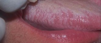
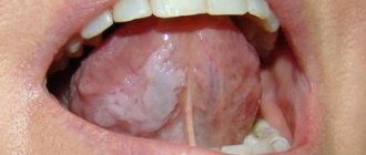
Clinical symptoms change significantly both in the presence of a banal infection and under the influence of treatment. Similar complaints and unclear development of the process often do not allow one to obtain a sufficient impression to determine a possible disease. In such cases, the diagnostic search begins with an objective assessment of the clinical picture, namely the appearance of the lesion elements. The subjective picture (complaints and description of anamnestic data) is reflected in detail in the medical record and occupies an important place in the diagnostic algorithm. As a specific example, consider the presence of white spots localized on the tongue, mucous membranes of the cheeks and lips: whitish areas with uneven borders (Fig. 1). The first step in the doctor’s thinking algorithm is to list diseases that have a similar visual picture. In this case, we should name candidiasis, lichen planus (LP), geographic tongue, desquamative glossitis, secondary syphilis.
After a thorough examination, returning to the analysis of complaints will allow you to link them to a specific disease. If no subjective sensations, paresthetic or painful, are noted, lichen planus, geographic tongue, or secondary syphilis can be assumed. Candidiasis and desquamative glossitis are characterized by pain when talking and eating food, especially spicy and salty food. Pain may appear with the development of the inflammatory-hyperemic form of LP or with syphilis in the event of a secondary infection. Geographic tongue can be combined with candidiasis, accompanied by pain. Thus, the presence of subjective complaints cannot serve as a pathognomonic sign of a specific disease. Finding out the onset of the disease from the anamnesis will allow you to take the next step in the diagnostic search. Candidiasis has a clear onset and accompanies common diseases or the use of medications that suppress the bacterial flora. Lichen planus can exist for quite a long time (months, years), but it appears in adulthood and is often caused by emotional stress. "Geographic tongue" is detected soon after birth and remains for many years. Desquamative glossitis (stomatitis) occurs and recurs, being associated with general diseases, most often pathology of the gastrointestinal tract (GIT). With syphilis, the rash of secondary elements of the lesion has a clear beginning and can appear against the background of preserved primary affect. The dynamics of the elements of damage have their own characteristic features. Plaque due to candidiasis occupies an increasingly larger area over time, moving to neighboring sections of the oral mucosa, and its quantity increases. With lichen planus, the picture is quite stable, only the clinical picture slowly and gradually increases or changes. “Geographical language” is distinguished by the “migration” of drawings. Desquamation of the mucosal epithelium in general diseases may increase over time, but the administration of anti-inflammatory drugs leads to improvement. With syphilis, the elements of the lesion gradually disappear, but on the tongue the picture of a “mowed meadow” is quite stable. A more detailed examination allows us to identify the features inherent in a particular disease. With candidal stomatitis, changes are observed in the mucous membrane of the entire oral cavity. When individual areas are affected, the disease, depending on the location, is called candidal glossitis, cheilitis, or tonsillitis. Acute candidiasis occurs in infants or weakened people (blood diseases, hypovitaminosis), as well as in persons receiving large doses of corticosteroids, cytostatics, and antibiotics. The patient is concerned about dry mouth and impaired taste. When eating food, especially salty, sour, spicy food, pain is noted. A characteristic sign of candidiasis is foamy saliva that collects in the retromolar region and on the back of the tongue. On the hyperemic, edematous mucous membrane of the cheeks, palate, gums, and tongue, whitish areas appear that merge to form a loose, “curdled” white coating (Fig. 2). After removing the plaque, a smooth, hyperemic mucous membrane is exposed.
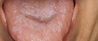
Rice. 1. The lesion elements are localized on the mucous membrane of the tongue. Rice. 2. Plaque on the mucous membrane with candidal stomatitis. Rice. 3. Chronic candidal glossitis. Rice. 4. Plaques with lichen planus on the tongue.
Subsequently, the plaque can become saturated with fibrin, taking on the appearance of a dense film of grayish or yellowish color, tightly attached to the surface of the epithelial layer. Pseudomembranous candidiasis, characteristic of HIV infection, develops. The initial symptoms of candidal glossitis may be pinpoint redness of the marginal zones and the tip of the tongue. With chronic candidal glossitis, small furrows with white deposits appear along the edges and at the bottom. The lesions are first detected at the base of the tongue, and then spread to the remaining parts, capturing its lateral surfaces. Against the background of atrophy of the filiform papillae of the tongue, a scant whitish coating is detected, which is not completely removed (Fig. 3). Candidiasis of the red border of the lips is manifested by dryness, hyperemia, swelling, and peeling. Painful erosions, small cracks, and thin gray scales may occur. Subjective sensations include tension and burning. The disease differs in its duration and relapses. The diagnosis of candidiasis is confirmed by the results of complex laboratory studies over time: microscopic, cultural (with determination of the type of fungus), and in some cases, histopathological. Fungi of the genus Candida are found in the form of yeast-like cells and pseudomycelium. The presence of pseudomycelium or chains of round or elongated budding cells upon microscopic examination of the material is considered sufficient to confirm the diagnosis. The predominance of yeast cells with single filaments of pseudomycelium indicates superficial candidiasis. The predominance of the filamentous form over the cellular form indicates the parasitic activity of the fungus and is more often detected in visceral lesions. Lichen planus (LP) can begin unnoticed, last for years, and be discovered accidentally when examining the mucous membrane by a specialist. In some cases, the abundance of rashes attracts the patient's attention. The etiology is not always determined. Often this is psycho-emotional stress, toxic-allergic effects. Elements of the lesion may first appear on the mucous membrane, then spread to the skin, or, conversely, a rash on the body precedes the lesion of the mucous membrane. The main element is always a papule: on the skin it is initially whitish, then pale pink, then reddish, lilac. Papules tend to cluster. The presence of skin rashes makes the diagnosis easier. On the red border of the lips, papules undergo keratinization, are connected by bridges, and form whitish areas in the form of individual raised nodules, bizarre patterns or merging areas of hyperkeratosis with uneven outlines. On the mucous membrane, PL is characterized by the presence of small whitish papules (up to 2 mm in diameter). The last few rise, which causes discomfort, a feeling of tightness or a feeling of roughness. Papules can acquire a grayish or pearlescent tint and tend to group together to form fancy patterns in the form of lace, mesh, or a “frosty pattern.” Favorite localization is the buccal mucosa along the line of closure of the teeth, retromolar region, tongue, gingival margin. On the tongue, the lesion elements can merge into plaques or be located in the form of circles, semi-arcs, or wavy lines (Fig. 4, 5). Papules in LP are painless; scraping does not remove the whitish surface. The exudative-hyperemic form is characterized by hyperemia, swelling of the mucous membrane, on which papules are located, forming a typical picture in the form of patterns, networks, arcs. Subjective sensations in the form of pain when eating food (hot, spicy, hard) are added. The bluish glow in Wood's rays of the lesion elements on the red border and the white glow on the mucous membrane makes it possible to differentiate the lesion elements in LP. The etiology of such a condition as “geographic tongue” has not been fully elucidated: neurotrophic disorders and diseases of the gastrointestinal tract are possible. In a number of cases, “geographical language” acts as a variant of the normal structure and is detected already in childhood. There may be no subjective sensations, and then the peculiarities of the appearance of the tongue are revealed during examination. Rarely, patients complain of tingling, burning, and paresthesia. Symptoms intensify with injuries to the mucous membrane, the development of Candida fungus, and the addition of a secondary infection. With the development of the “geographic tongue” picture against the background of a general disease, the process of desquamation begins with the appearance of a small area of turbidity of the epithelium; in the center, the upper layers of the keratinized epithelium are peeled off, revealing a pink, smooth area that quickly grows along the periphery. Multiple or single lesions reach a diameter of 1-2 cm, having the shape of spots, rings, half-rings, the boundaries merge with the surrounding mucous membrane. In the center, normal keratinization of the filiform papillae begins. The lesions are layered, new ones appear against the background of the old ones, which gives the surface of the tongue the appearance of a geographical map (Fig. 1). Minor keratosis may appear in the form of whitish stripes. In some cases, pigmentation of these areas occurs (Fig. 6). The process usually does not extend to the lower surface of the tongue. A similar picture rarely occurs simultaneously on the lips, cheeks, and gums. A constant feature is the preservation of filiform papillae of the tongue.
Rice. 5. Lichen planus in the form of a lace pattern. Rice. 6. “Geographic tongue” is combined with a deep median sulcus. Rice. 7. Desquamation of the epithelium is combined with plaque during gastritis. Rice. 8. Picture of a “mowed meadow” with syphilis.
Cytological examination reveals epithelial cells with keratohyalin inclusions. Desquamation of the epithelium, combined with plaque, is characteristic of diseases of the gastrointestinal tract (GIT). In such cases, discomfort and pain appear in the oral cavity, especially during meals. The tongue may be swollen, and then teeth marks can be identified on it. Cracks, erosions, and areas of increased desquamation form on the tongue, cheeks, and lips (Fig. 7). Gastritis with reduced secretion is characterized by dry mouth, complaints of burning, pain in the tongue, especially from spicy, hot foods, and a metallic taste. Against the background of atrophy of the filiform papillae of the tongue, desquamation of the epithelium is characteristic. The filiform papillae are smoothed, the mushroom-shaped papillae appear hypertrophied. With atrophic gastritis, atrophic glossitis develops. Characterized by burning and pain in the tongue. The tongue is pale pink, partially coated, the filiform papillae are atrophied. With a gastric ulcer, plaque on the tongue may be more or less abundant and pigmented, but can be easily removed. There may be complaints of a burning sensation (the tongue feels as if it is scalded or sprinkled with pepper), pain and an increase in the size of the tongue due to swelling. There are teeth imprints on the side surfaces. Independent glossitis may develop: “geographical”, “black hairy” tongue. Fungal stomatitis is often associated. A course of treatment for a general disease leads to an improvement in the condition of the oral cavity, but does not exclude relapses of desquamative stomatitis (glossitis). The secondary period of syphilis in the absence of treatment manifests itself after 6-8 weeks as a fresh secondary one, and then its relapses can be repeated for 3-5 or more years, lasting 1.5-2 months and characterized by more or less profuse rashes. About 50% of patients have manifestations in the oral cavity, and this is often the only localization of the lesion elements. The most common lesions found are papular rashes. Favorite localizations are tonsils, palatine arches, cheeks (along the line of teeth closure), tongue. Red papules are small at first, then grow to several millimeters and become covered with a peculiar grayish coating. After scraping with a spatula, flesh-red erosion is exposed. Papules tend to spontaneously erode. The addition of a secondary infection leads to pain.
When localized on the back of the tongue, syphilitic papules acquire the characteristic pattern of a “sloping meadow” - clearly defined oval areas with a smooth surface. The latter is formed due to atrophy of the filiform papillae (Fig. 8). Clinical diagnosis of syphilis is based on the characteristic elements of the lesion with mandatory confirmation by the results of laboratory tests to identify the pathogen or specific reactions.
Conclusion Difficulties in diagnosing diseases affecting the oral mucosa are associated with the lack of clear differences between subjective and objective symptoms. In such cases, the dentist can use a diagnostic search algorithm (without violating the rules and procedure for filling out medical documentation). After receiving the entire amount of information, he consistently excludes diseases that do not have characteristic features inherent in the clinical manifestations of a given patient.
Oral cavity examination methods
Methods for examining the oral cavity come down, first of all, to a thorough examination using directional (preferably shadowless) lighting and special instruments (see Dental Instruments) - a spatula, wide hooks for retracting the cheeks, lips, tongue and stomatol. mirrors for inspecting hard-to-reach areas. Sometimes, to identify luminescent compounds in the oral mucosa (see Luminescence), an inspection of the oral cavity is performed under UV light. During the examination, pay attention to the presence of bad breath. With the help of palpation, the mobility, density, consistency and soreness of various parts of the mucous membrane and patol are determined. formations.
To study the organs surrounding the oral cavity, various X-ray diagnostic methods are used (see). Research methods such as ultrasonic echolocation (see Ultrasound diagnostics) and thermography (see) are also used. According to indications, Citol is produced. examination of swabs and prints from pathologically changed areas of the mucous membrane (see Cytological examination), as well as the study of the microflora of the oral cavity. According to strict indications, a biopsy is performed (see).
In some cases, there is a need to conduct immunological and biochemical studies, as well as to determine various types of sensitivity: tactile, pain, temperature, taste (see Esthesiometry).
See also Patient examination, dental examination.
Pathology
Pathologies of the oral cavity include malformations, diseases of the oral mucosa, innervation disorders, diseases of the organs surrounding the oral cavity, and tumors.
Developmental defects
may refer to congenital defects of the lips (see Lips), palate (see), jaws (see), tongue (see); Congenital facial clefts are rare (see).
Diseases of the oral mucosa
Diseases of the oral cavity include, first of all, lesions of its mucous membrane, characterized by a variety of morphols. violations and wedge, manifestations, which often presents serious difficulties in differential diagnosis. There are several main groups of diseases of the oral mucosa.
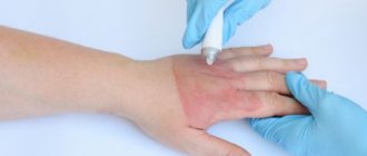
Traumatic damage to the oral mucosa can be caused by mechanical, chemical, thermal, and radiation factors. The severity and duration of the course depend on the size and depth of the lesion, however, wounds and damage to the mucous membrane heal faster and are less likely to be accompanied by complications than similar skin damage. Long-term exposure to irritating factors can lead to the formation of traumatic erosions, hron. ulcerations, decubital ulcers. The cause of mechanical trauma can be the crowns of incorrectly erupted or displaced teeth, sharp edges of carious cavities, incorrectly applied fillings and artificial crowns, uneven edges of dentures, their clasps, tartar deposited on the surface of the teeth (see). Irritation and damage to the mucous membrane can occur as a result of ingestion of excessively hot, hot, spicy food, strong alcoholic drinks, especially often smoking, as well as as a result of certain traditional bad habits: chewing tobacco, betel leaves, etc. Often the effect of chronic irritants and traumatic factors leads to disruption of the process of keratinization of the epithelium of the mucous membrane, hyperkeratosis (see), leukoplakia (see).
Inflammatory diseases of the oral mucosa - stomatitis (see) are distinguished by the location of the lesion, etiology, morphol. changes and wedge, flow. According to localization, inflammation of the mucous membrane of the lips, their red border is distinguished - cheilitis (see), tongue - glossitis (see), gums - gingivitis (see).
Allergic reactions (see Allergy) are a relatively common cause of damage to the oral mucosa. Some of them are classified as infectious-allergic, for example, chronic recurrent aphthous stomatitis; others are caused by chemicals, especially drugs, or are local manifestations of general allergic reactions.
The oral mucosa can react to various patols. processes and functional disorders in many body systems. Its characteristic changes are sometimes the earliest symptoms of diseases of the digestive, excretory, circulatory systems, blood diseases, as well as hypovitaminosis and many inf. diseases. Characteristic changes in the oral cavity are accompanied by certain dermatoses (see). Changes in the oral mucosa are important for diagnosing professional and household chronic intoxication with various chemicals, for example, heavy metals (See Mercury, Lead).
Tumors of the oral cavity can develop both from the mucous membrane and spread from deeper tissue structures and organs. Of the benign tumors, the most common are papillomas (see Papilloma, papillomatosis), fibromas (see Fibroma), cystic formations of small salivary glands located in the thickness of the walls of the oral cavity, the so-called. mixed tumors of the glands (see Mixed tumors). Vascular tumors - hemangiomas (see Hemangioma), much less often lymphangiomas (see Lymphangioma) can be localized in different parts of the oral cavity.
Among malignant tumors, cancer is of great importance in the pathology of the oral cavity. Cancer of the oral cavity, including cancer of the lips (see Lips) and tongue (see), accounts for about 10% of all cancers. It is generally accepted that cancerous lesions often develop in areas of the mucous membrane that have chronic. damage, ulceration, cracks, as well as in areas affected by hyperkeratosis, leukoplakia, and certain other so-called. precancerous diseases (see Precancerous diseases). Timely detection and elimination of precancerous diseases of the oral cavity is the most important part of cancer prevention.
Disturbances in the innervation of certain areas of the oral cavity can manifest themselves in the form of loss of sensitivity (analgesia), the occurrence of distorted and unpleasant sensations (paresthesia) and various pain syndromes associated with neuritis or neuralgia of individual branches or branches of the nerves involved in the innervation of the oral cavity and its organs. One of the typical and relatively common types of such a disorder is glossalgia (see), manifested in the form of pain or a burning sensation in the tongue.
Diseases of other organs related to the oral cavity
Along with diseases of the mucous membrane, one of the most common types of oral pathology are dental diseases: caries (see Dental caries), pulpitis (see), periodontitis (see), periodontal disease (see), as well as dental anomalies (see .), dentition, bite (see).
Severe forms of odontogenic inflammatory processes are periostitis and osteomyelitis of the jaws (see), abscesses (see Abscess) and phlegmon of the surrounding soft tissues (see Phlegmon), in particular phlegmon of the floor of the mouth (diffused purulent inflammation of the tissue of the intermuscular and interfascial spaces between the body of the mandible and the hyoid bone), as well as Ludwig's angina (see Ludwig's angina). Treatment of phlegmon consists of opening through wide incisions all possible areas of accumulation of pus and draining them in combination with intensive measures of general anti-inflammatory therapy.
Among diseases of other organs associated with the oral cavity, it should be noted diseases of the salivary glands that affect the condition of the oral mucosa and disrupt the functions of the oral cavity (see Xerostomia, Sialadenitis, Sialolithiasis).
For a number of diseases of the oral mucosa of an inflammatory or necrotic nature (ulcerative necrotic stomatitis, gingivitis, etc.), dental diseases (caries, periodontal disease, pulpitis, etc.), chronic. tonsillitis, for diseases of the upper respiratory tract (ozena, decaying tumor), lungs (bronchiectasis), gastrointestinal tract. tract (anacid gastritis, esophageal diverticulum), metabolic disorders (diabetes mellitus, scurvy, etc.), bad breath (foetor ex ore) may be observed, to eliminate which treatment of the underlying disease is necessary.
Treatment
For purulent processes on the Lip (furuncle, carbuncle), treatment is mainly conservative; you should not squeeze out the so-called. rods. Good results are obtained by using local novocaine blockade with antibiotics with simultaneous intramuscular administration of broad-spectrum antibiotics. In the first stage of inflammation, during the period of infiltration, radiotherapy at 120 kV, a 1-3 mm Al filter, a field that covers normal tissues surrounding the infiltrate by 1-1.5 cm, with a single dose of 15-25 r daily or every other day up to a total dose of 75-125 r. Under the influence of radiation, the infiltrate disappears, surgical intervention is not required. Surgical treatment is indicated for a formed abscess (see Carbuncle, Furuncle).
Treatment of malignant tumors can be divided into treatment of the primary tumor and regional metastases.
To treat the primary tumor, radiation therapy or a combined method is used (in the first stage - radiation therapy, in the second - wide excision with primary plastic surgery). Treatment of regional metastases is carried out mainly by surgery.
Radiation therapy for cancer of G. is carried out using the methods of interstitial gamma therapy (see), close-focus radiotherapy (see), electronic therapy (see), and, less often, application gamma therapy.
For the treatment of patients with stage I–II cancer, close-focus radiotherapy and interstitial gamma therapy are indicated. At stage III, interstitial gamma therapy and electron therapy have an advantage. For stage IV G. cancer, combined radiation therapy is indicated: remote gamma therapy or electron therapy, followed by the use of close-focus radiotherapy or interstitial gamma therapy. When the mucous membrane and skin of the mouth are affected, when the tumor is localized in the corners of the mouth, as well as when cancer recurs, the interstitial method has an advantage.
A contraindication for radiation therapy is the presence of a concomitant inflammatory process, upon elimination of which radiation therapy can be carried out. A contraindication for interstitial gamma therapy and close-focus radiotherapy is also the spread of the tumor to bone tissue and the inability to determine its boundaries, and in case of relapses, significant radiation changes in the surrounding normal tissues.
For close-focus radiotherapy, a single dose is 400-500 rad, the total dose to the lesion is 6000-6500 rad; irradiation field no more than 25 cm2.
In the interstitial method, needles with 226Ra, 60Co are used; The most convenient are nylon threads with 60Co granules. Radioactive drugs are administered after local anesthesia with 0.25% novocaine solution. Irradiation is continuous for 6-7 days. The total focal dose is 5000-7000 rad at a dose rate of 30-40 rad/hour.
For electronic therapy, Betatron type devices with radiation energy of 8-15 MeV are used. A single dose is 400 rad, the total dose is 5000-7000 rad, if; used as the only method. When combined with the interstitial method, the dose from electronic therapy is reduced.
The application method using 60Co preparations allows for fractional treatment with a daily dose of 500-600 rads and a total dose of 5000-6500 rads.
During radiation therapy, protection of the alveolar part of the lower jaw is required; the edges are provided with plexiglass or methyl methacrylate gaskets between the jaw and the jaw bone.
In stage I cancer of the lower cervix, a permanent cure is achieved in 95-96% of cases; Regional lymph nodes are not removed. Radiation therapy provides a high percentage of radical cure, better cosmetic and functional results compared to the surgical method, and fewer cases of relapses and metastases.
In stages II-IV of cancer, when curing the primary tumor, even in the absence of enlarged lymph nodes, an upper cervical excision operation should be performed, in which not only the submandibular and submental lymph nodes, but also the deep cervical lymph nodes located in the bifurcation area are removed carotid artery. In the presence of clinically significant regional metastases, preoperative external gamma or electron therapy with conventional dose fractionation and a total dose of 4000–4500 rad is indicated. The operation is performed after 2-3 weeks. after completion of radiation therapy.
Lip operations
taken to treat wounds, for purulent processes, for the treatment of tumors, etc.; A special place is occupied by operations on children and plastic surgeries.
Primary surgical treatment of wounds of G. should be performed taking into account functional and cosmetic requirements. Excision of tissue should be minimal and only tissue that is obviously non-viable and crushed. When applying layer-by-layer sutures, it is imperative to restore the continuity of the orbicularis oris muscle. The suture should be applied especially carefully to the skin and red border of the lips. In case of injury with a large defect in the tissue of the lips, when the edges of the wound cannot be sutured without tension, primary plastic surgery should be performed using tissue from areas of the face adjacent to the defect.
If the frenulum is thick and short, limiting G.’s mobility, it is excised ( frenectomy
) . To avoid scar formation, it is better to make a middle incision along the frenulum and use opposing triangular flaps.
With the so-called the double lip is surgically removed; excess submucosal tissue and mucous glands and fix the mucous membrane to the G muscle.
The retention cyst is removed with suturing of the mucous membrane. A mixed tumor should be removed along with the capsule and the mucous membrane covering it. The papilloma is excised with a small area of adjacent tissue. For small-sized hemangioma and lymphangioma, they are excised. With diffuse hemangioma, it can be reduced by introducing 70% alcohol into it to produce tissue sclerosis. For keratoacanthoma, they resort to either excision or close-focus radiotherapy.
Operations
Small-scale surgical interventions in the oral cavity are performed, as a rule, by dental surgeons, most often on an outpatient basis. Before any operation in the oral cavity, the oral cavity is sanitized (see).
The most common are tooth extraction operations (see Tooth extraction), as well as interventions associated with diseases of the periodontal tissues and odontogenic processes. They are usually performed under local (in the lower jaw, mainly conduction) anesthesia (see Local anesthesia, maxillofacial area). Opening of gingival abscesses and purulent foci during periostitis is carried out using incisions to the bone followed by drainage.
Incisions for cysts (see Dental cyst), benign neoplasms, and pathologically altered areas of the mucous membrane are made within unchanged tissues, followed by suturing with catgut. Wound suturing for oral injuries is performed tightly. Features of surgical treatment of wounds for wounds, including gunshots, affecting the oral cavity - see Face, Jaws.
More extensive interventions in the oral cavity are performed in an inpatient setting under local anesthesia with premedication or general anesthesia. Plastic surgery is performed in the presence of congenital malformations (cleft lip and palate, “double lip”, shortened frenulum of the upper lip and tongue, etc.), as well as for the consequences of injuries and diseases (scars, defects).
In cases of cicatricial deformities in the area of the corners of the mouth, to eliminate the narrowing of the oral fissure, the so-called. microstomy, the edges of the oral fissure with their end-to-end scar change are dissected and epithelialized, everting the mucous membrane from the cheek (Evdokimov’s method). If the strip of the red border is preserved, it is cut off with a through incision, keeping it in the form of a bridge between the upper and lower lips, and then, after dissecting the scars and tissues, the cheeks are pulled up to the required level, where they are secured with sutures, thus forming a new corner of the mouth (Vasiliev’s method).
It is very rare to resort to plastic surgery for an excessively wide mouth gap, the so-called. macrostomy, resulting from a unilateral transverse cleft of the face of a congenital nature (see Face, malformations).
In the postoperative period, careful hygienic care of the oral cavity is necessary (excessive rinsing of the mouth, rinsing), as well as the administration of liquid or softened food, introducing it through a sippy cup if chewing is impossible.
Standards for a healthy oral cavity
Home / Articles / Standards for a healthy oral cavity
How is oral health determined? A healthy oral cavity is the absence of diseases, inflammations and pathologies. Orthodontist Farida Yusifovna Gasimova talks about the health standards of the oral cavity and dental system.
— Farida Yusifovna, how is dental health determined?
— Disorders in the dental system provoke malfunctions in the joints and muscles. Fixation of the lower jaw is carried out with the help of the muscles of the head and neck; the chewing process occurs when the temporal and masticatory muscles work. If the chewing apparatus is functioning correctly and the joints and muscles are anatomically normal, there is a distance (2 mm) between the teeth of the upper and lower jaws. Disturbances in the bite occur with excessive muscle tone in a calm state, which affects not only the teeth, but also general well-being: the patient complains of frequent headaches, pain in the neck muscles, in the back, there is pinching of the facial muscles, teeth are subject to wear, chips appear at the ends, and a wedge-shaped defect is often diagnosed. Disturbances in the dental system can be of a different nature; based on external symptoms, one can more or less understand that it is necessary to consult an orthodontist. If the patient notices stiffness when moving the lower jaw, jamming of the jaw, characteristic clicks when it moves (crunching, clicking) - this indicates problems with the temporomandibular joint. The action of the TMJ (temporomandibular joint) is aimed at ensuring normal movement of the lower jaw. The temporomandibular joint looks like a dimple with a head, with an articular disc placed between them. The absence of disturbances is manifested in the form of the location of the head in the fossa (resting state). When the jaw moves, the head is displaced by the articular disc. If there are disorders, the articular disc is displaced, the bite is disturbed and the jaw moves incorrectly. Dysfunction of the TMJ (temporomandibular joint) requires complex treatment from doctors of various specialties, including a gnathologist, an orthodontist, and a neuromuscular dentist. The condition of the dental system is significantly influenced by the patient’s spine and posture. If posture is impaired, the muscles of the face, neck, and back cannot work correctly, and this fact, in turn, affects the normal functioning of the lower jaw. The displacement of the lower jaw causes disruptions not only in the functioning of the dental system, but also in the body as a whole. An anatomically correct bite is not an indicator of a healthy dental system, as many patients think. Just like white, straight teeth. The dentofacial system generally cannot be characterized by any one of its organs, since, as I have already said, a violation in any of its components affects the entire system. The condition of the dental system is directly related to the work of the ear organs, temporomandibular joints, neck and head muscles. The psycho-emotional state of the patient also plays an important role. If pain in the neck and back periodically appears, this already indicates an existing disorder, including in the dental system. Therefore, only a dentist orthodontist in Anapa can determine its condition.
— What is the dental system?
— The dentofacial system is a complex apparatus in which the organs are anatomically united. The organs included in the dental system are bones (palatine, zygomatic, nasal, jaw), teeth, temporomandibular joints (temporomandibular joints, lips, cheeks, tongue, soft and hard palate, salivary glands, masticatory and facial muscles. Functioning separately and together, all these organs give us the opportunity to breathe, talk and eat food. I want to emphasize that in the dental system there are no “important” and “minor” organs, each individual component of the system plays its role. The growth and formation of the dental system begins in changes in the womb and throughout a person’s life. From childhood to final adulthood, modifications of the dental system are clearly noticeable, since during this period the bite changes, individual features of the cheekbones, tongue, and lips are formed. Then the changes are little noticeable, but they continue to occur. When there is a process of active formation (childhood), the Anapa orthodontist dentist can quickly correct the incorrect growth trend to an anatomically correct one, direct the growth of teeth and bones, so to speak, in the right direction. You should not wait until the jaw is fully formed (adulthood) for orthodontic treatment; in children, therapy is faster, easier, and cheaper. Dentistry Millennium Anapa Mayakovsky offers patients orthodontic services. It is also important to note the fact that a disorder in one organ inevitably affects the functioning of the entire system, therefore, if the pathology is not corrected during the period of formation and growth of the jaws, it can develop into a severe disorder in adulthood. It is necessary to undergo an examination by an orthodontist during the period of 5-7 years, which will exclude or identify a tendency to disrupt the development of the dentofacial system. A pediatric orthodontist in Anapa conducts a visual examination and diagnosis using special equipment. I repeat, it is much easier to carry out correction in a child than long-term therapy in an adult patient.
— Farida Yusifovna, how are teeth treated today?
— Treatment of the dental system must be carried out comprehensively, with mandatory visits to specialized specialists. Anapa Mayakovsky Dentistry Millennium dental treatment, in the clinic you can get a consultation with one of the best dental specialists in the city and undergo a full diagnosis of the dental system. Depending on the patient’s clinical picture, the orthodontist in Anapa prescribes consultations with doctors and computed tomography. Therapy of the dental system is always carried out according to an individual program; in case of violations, it is impossible to treat everyone according to the same scheme. Electromyography (myography) is used to identify dysfunction of the masticatory and temporal muscles and determine the affected nerve areas. Using electromyography, it is possible to determine the causes of paralysis of an individual muscle. The principle of operation of the myograph is that it records changes in the bioelectrical activity of muscles using special sensors attached to the facial muscles and connected to the myograph. Therapy of the dental system includes treatment with an occlusal guard and a splint (a removable device for correcting the bite). With the help of a mouthguard, pain in the TMJ, pinching of the lower jaw, headaches and other disorders are eliminated, the action of the occlusal splint is aimed at restoring the natural position of the muscles during the functioning of the jaw, with the help of the splint the symmetry of the face and anatomically correct muscle tone are restored. Another technique that deserves attention is electrical neurostimulation (TENS), the effect of which is aimed at relaxing the masticatory muscles that are in hypertonicity. This method effectively eliminates muscle spasms, saturates tissue cells with oxygen, ensures the correct position of the lower jaw, removes lactic acid, and restores proper muscle function. TENS therapy is carried out using a current that is passed through the patient's skin; the entire procedure takes about one hour. The facebow is used to accurately analyze the individual position of the jaws in three dimensions in order to then produce a prosthetic structure (veneers, dentures or crowns). The use of this mechanical device in dentistry can significantly reduce errors in the design of orthopedic devices. Making a structure for the dental system is a rather complex task that requires maximum attention, experience and professionalism; not only the individual characteristics of the position of the bones are taken into account, but also the dynamics of movement. When making orthopedic structures, it is very important to match the central occlusion and jaw relationship. When conducting therapy, dentists set themselves the priority tasks of restoring proper functionality, and only then the aesthetic factor. Of course, most patients are primarily interested in the beauty of a smile, but if beautiful teeth are not capable of fully chewing food, then, of course, it is much more important to recreate functionality.
Comprehensive hygiene for 2500 rubles! Air Flow 1000p for patients with braces!
A unique promotion for our regular patients! A set of hygienic procedures (removal of tartar and plaque using Air Flow and ultrasound , scaling, polishing, fluoridation) for regular patients once every 6 months for RUB 3,000!
Air Flow and polishing for only RUR 1,500 for patients with braces!