Content:
- Why isn't every spot a black tooth decay?
- When there is black caries, but it is still not treated
- How to deal with little black caries
- When black caries still needs to be filled?
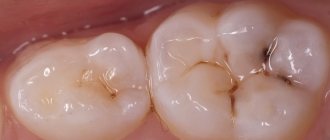
Sooner or later, small dots or dark-colored depressions appear on the teeth of any person. They are usually perceived as caries. But, when visiting a dentist, the patient may be told that he does not need to treat his teeth. Why is this happening? Is it true that not every black spot on the crown of a tooth is a cavity?
Causes of darkening of enamel
Discoloration of enamel and dentin can be caused by:
- carious lesion,
- pulp necrosis,
- long-term use of tetracycline antibiotics in high dosages,
- violation of the acid-base balance in the oral cavity,
- overdose of fluoride or zinc,
- smoking,
- some diseases of the gastrointestinal tract,
- improper filling,
- excessive consumption of coloring foods and drinks - black coffee, strong tea, pomegranate and grape juice, soy sauce, blueberries and blackberries.
To return the enamel to its natural color, you need to make an appointment with a dentist. The doctor will determine why the crown of the tooth has darkened and will suggest the optimal restoration method, taking into account the age and health characteristics of the patient.
Why isn't every spot a black tooth decay?
There is no need to think that dentists are being cunning, wanting to deliberately turn an initial carious cavity into mid- or deep-stage caries.
After all, the more complex the problem, the more expensive its treatment. No doctor does this. It’s just that the identified violation has a completely different origin. In reality, dark spots can represent:
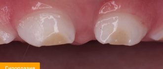
- Consequence of hypoplasia. This is a dental disorder in which, during the formation of a tooth, its crown part is underdeveloped. The disease is a consequence of metabolic disorders in the buds of teeth. It can also develop due to improper metabolism of proteins and minerals in the fetal body during its intrauterine development. Rickets, somatic pathologies, diseases of the gastrointestinal tract, severe infectious diseases - all these are also factors that provoke hypoplasia.
- The result of fluorosis. This disease occurs due to the constant and prolonged ingestion of large amounts of fluoride into the body. First, whitish spots form on the crowns, and then depressions resembling black caries. During examination, the doctor records destructive and erosive changes in the enamel.
- Erosion of teeth. Another type of non-carious lesion of tooth enamel. Defects occur on the outer layer of the crown and can even affect the dentin. Erosive defects are most often localized symmetrically. They provide an unsightly cosmetic effect and therefore require urgent correction.
In many non-carious pathologies, it is possible to improve the aesthetics of a smile through remineralization. The patient is prescribed vitamin and mineral complexes, calcium and phosphorus supplements, and local applications. Electrophoresis is used in many clinical situations.
Only with voluminous cavities reminiscent of black caries do they resort to filling material. If the damage is very severe (which happens when a person puts off visiting the dentist for a long time), even prosthetics may be required.
It is important to understand that caries is always a cavity of microbial origin. That is why it has to be drilled out to remove all damaged tissue and prevent further spread of pathogens. From this position, not every even point on the tooth requires the use of a drill.
Symptoms and diagnosis
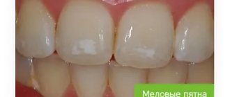
At the initial stage of the disease, caries, as a rule, does not manifest itself in any way. In most cases, the tooth does not hurt, and only the appearance of a stain on the enamel, which most often has a round or oval shape, can cause concern. Its shade can be chalky white or yellowish.
Diagnosing a stain on the enamel surface is not always easy, even in visible areas, if the color changes are minor. To identify this stage of caries, in addition to examination and probing, the following methods are used:
- Drying. After removing moisture from the surface of the tooth, stains appear more clearly on it.
- Vital coloring. Special dyes are applied to the enamel, which are absorbed well even by minimally caries-affected enamel.
- Luminescent method. When exposed to ultraviolet radiation, a shadow forms in the area of the carious spot.
When there is black caries, but it is still not treated
Many people have encountered a situation where a doctor, during an examination, confirmed the presence of the initial stage of caries, but refused to treat it according to the standard regimen. Why is this happening? The thing is that during drilling of dentin, much more hard tissue is damaged than it seems at first glance. But it is impossible to install a seal without drilling.
Trying to avoid unnecessary destruction of the crown, the doctor does not want to drill into black caries at the spot stage. Very often its dimensions do not change at all for a long time. The spot itself does not cause a person any discomfort. If you brush your teeth thoroughly after each meal, use high-quality toothpaste and remineralizing dental gel, you can maintain caries at its original size for several years, that is, avoid its increase.
Therefore, if the dentist says that there is no need to touch the tooth yet, you should listen to his words. This is a way to preserve hard tissues without changing their volume for the coming years.
Treatment of dental granuloma –
Treatment for dental granuloma is usually only therapeutic. Treatment consists of mechanical treatment of the root canals, in most cases - temporary filling of the root canals with special medicinal pastes based on calcium hydroxide, after which after 2-3 weeks it will be possible to permanently fill the root canals with gutta-percha, as well as put a permanent filling. Below we will talk in more detail about how exactly this treatment is carried out.
Surgical treatment of tooth root granuloma may be required only in a few cases. For example, the operation of resection of the root apex may be indicated - 1) if the root canals are obstructed, 2) if it is impossible to unseal previously poorly filled root canals in order to treat the source of inflammation at the root apex, 3) if there is an artificial crown on the tooth, 4) if in A pin is installed in the root canal, an attempt to remove it may lead to a fracture of the tooth root.
Therapeutic treatment strategy –
- If the root canals have not been previously filled, it is necessary to drill out all carious tissues and old fillings, after which mechanical treatment of the root canals is carried out. After their expansion and antiseptic treatment, the strategy for further treatment may depend on the size of the granuloma.
If the tooth granuloma is small in size (up to 2-3 mm), then the root canal of such a tooth can, in principle, be filled immediately. If the size of the granuloma is from 3 to 5 mm, then a special therapeutic material based on calcium hydroxide is left in the root canals, which will allow the granuloma to shrink or even disappear completely.Temporary canal filling is carried out for a short period of time (on average 2-3 weeks). After this period, a control X-ray is taken, and if the granuloma has disappeared or decreased, the root canals are filled on a permanent basis, and a permanent filling is placed on the tooth. Algorithm for the therapeutic treatment of granulomas more than 3 mm in diameter, cystogranulomas and cysts (detailed descriptions appear when opening the photo) –
- If the root canals in this tooth are sealed (Fig. 7-11) - in this case, the root canals must first be unsealed, and then the treatment sequence corresponds to the previous option.
If such a tooth has an artificial crown, then usually they are first removed and the tooth is treated. And it must be said that in this case the crown will have to be made again. If you do not want to spend money on replacing the crown and filling the root canals, then in principle it is possible to do without it. A surgical root resection operation will help you with this, which means that through a small incision in the gum, the apex of the root with the unfilled part of the root canal and with a granuloma attached to the apex of the root will be cut off from the root.
Emergency care for purulent inflammation -
If you have a dental granuloma, then an exacerbation of the chronic process can lead to the development of purulent inflammation.
If the pus is localized inside the granuloma, then relief from acute pain is possible only after opening the tooth and unsealing the root canals, which will allow the pus to be released through them. How the outflow of pus occurs through the root canals - see video 1 below. But if there is significant swelling of the gums or cheeks, which indicates the release of pus under the periosteum or mucous membrane of the oral cavity, an incision will be required to release the pus. How to make a gum incision - see video 2.
Important: in some cases, a tooth with granulomas may need to be removed (for example, when the tooth crown is severely damaged and cannot be restored). In this case, it is very important that when removing a tooth, the dental surgeon must remove the granuloma from the tooth socket. We have already said above that in most cases they are tightly attached to the apex of the root, and therefore come out along with the tooth. However, in some cases, the granuloma remains in the socket, and then inflammation of the socket of the extracted tooth occurs (alveolitis).
What does a granuloma look like when scraped out of a socket after tooth extraction?
Treatment of dental granuloma with antibiotics -
It is impossible to treat dental granuloma with antibiotics. They can be prescribed only in case of exacerbation of the process (if purulent inflammation occurs) - in parallel with dental treatment carried out by the dentist. The fact is that it is impossible to sanitize infected root canals by simply taking antibiotics, which means the source of infection and inflammation will not disappear. By taking antibiotics, you can only achieve a temporary subsidence of the inflammatory process, but the granuloma itself will not even decrease in size. We hope that our article was useful to you!
Sources:
1. Dental education of the author of the article, 2. Based on personal experience as a dentist, 3. National Library of Medicine (USA), 4. “Therapeutic dentistry: Textbook” (Borovsky E.), 5. “Practical therapeutic dentistry” ( Nikolaev A.).
How to deal with little black caries
To prevent the cavity from turning into a large hole, you need to perform remineralization in the dentist's office. To do this, the doctor applies a special composition to the teeth, which quickly penetrates into the deep layers of the damaged units and, as it were, nourishes them from the inside. As a result, the teeth become stronger and more durable, and the rapid spread of the carious process is temporarily blocked.
In case of serious damage, the dentist can give the client a mouth guard and a special remineralizing composition. You need to use all this at night - before going to bed, put the product in a mouth guard and put it on your teeth. Remove in the morning. Repeat the procedure the number of times indicated by the doctor.
Prevention
Thorough oral hygiene is the key to dental health! A small black dot will not appear on the tooth unless the patient uses metal objects instead of a toothpick. There should be no microscopic damage to the gums and enamel, then infections will not appear. Since teeth are living organs, mineral and vitamin deficiency should be avoided. To do this, balance your diet, and if necessary, take complexes with vitamin and mineral supplements. Visit the dentist regularly. Visits to the doctor at least once every six months will allow you to identify diseases in a timely manner.
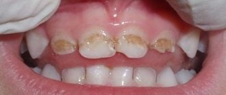
When black caries still needs to be filled?
You need to clean a black spot using a drill if:
- the patient complains of increased sensitivity to sour, salty and sweet foods, temperature changes;
- when there is mechanical impact on the damaged crown, pain occurs;
- the hole is growing in size too quickly;
- the speck is located in the area of the neck of the tooth;
- there was an unpleasant odor from the mouth;
- The black defect is localized on the incisors and spoils the smile.
In all these cases, the fight against carious lesions is carried out according to the classical scheme. The patient is injected with an anesthetic, after which the carious lesion is cleaned from altered tissues using a drill. The cleaned area is treated with an antiseptic and filled with a filling solution. The latter is dried with a lamp and then sanded to achieve the correct shape and perfect smoothness. As a result, the unit begins to look aesthetically pleasing, and its service life increases significantly.
Methods for solving the problem
The method of tooth restoration is chosen taking into account:
- causes of darkening of enamel and dentin,
- the patient has chronic diseases.
The following techniques are used in modern dentistry.
- Removing plaque and tartar. Professional oral hygiene can make the enamel 1-2 shades lighter and restore its smoothness.
- Endobleaching. It is carried out when there is discoloration of one or more pulpless teeth, as well as poorly performed prosthetics. The brightening gel is injected directly into the tooth canals.
- Restoration of the crown part of the tooth with composites and other materials. The doctor removes damaged tissue, treats the cavity, and restores the crown.
- Microprosthetics. Indicated in difficult cases when it is impossible to restore a darkened and damaged crown using other methods. It involves installing a crown made of metal-ceramics and other materials.
In rare cases, tooth extraction is recommended. It is shown if:
- the roots are damaged, there is a large crack on the crown,
- there is severe loosening of the tooth, inflammation of the gums,
- the inflammatory process affects not only soft tissues, but also the jaw bone.
After tooth extraction, a crown or implant can be installed.