Salivary glands
[
glandulae oris
(PNA, JNA, BNA);
synonym: glands of the mouth
, T.] - digestive glands that secrete into the oral cavity a specific secretion that is part of saliva.
There are large - parotid, submandibular, sublingual and small salivary glands - buccal, molar, labial, lingual, hard and soft palate (Fig. 1). Rice. 1. Schematic representation of the localization of the main human salivary glands: 1 - molar glands; 2 - buccal glands: 3 - labial glands; 4 - anterior lingual gland; 5 - sublingual gland; 6 - submandibular gland; 7 - parotid gland; 8 - accessory parotid gland
Comparative anatomy and embryology
In animals that live in water, the glands of the mouth are poorly developed and are represented by simple glands that produce mucus. In terrestrial animals, due to the need to moisturize the oral mucosa and wet food, the salivary glands are more developed. Amphibians have mucous labial, palatine, lingual and premaxillary glands. In reptiles, in addition, sublingual glands appear; in birds, the sublingual and so-called glands are well developed. angular glands. In mammals (except cetaceans), in addition to numerous small salivary glands, large salivary glands appear, located outside the oral cavity.
In human embryogenesis, all glands of the mouth arise as a result of the ingrowth of cellular elements of the stratified squamous epithelium of the mucous membrane into the underlying mesenchyme. The minor salivary glands develop from the 3rd month of embryonic development; by the 5th month, excretory ducts are formed and the glands begin to function. Large S. develop from epithelial strands growing into the underlying mesenchyme, which during the growth process divide and form branching ducts and end sections. The formation of the parotid gland occurs at the 6th week, and the submandibular gland at the end of the 6th week. embryonic development. At 7-8 weeks. Several anlages of the sublingual glands appear, from which independent glands are formed; their terminal sections are united by a common capsule and open into the oral cavity with 10-12 separate openings.
Physical properties and composition of saliva
Biological fluid in a healthy person has a number of physical and chemical properties. They are presented in the table.
Table 1. Normal characteristics of saliva.
| Index | Characteristic |
| Transparency | Transparent, minor inclusions of air, pieces of food. |
| Density | Slightly higher than the density of water, depending on the composition - from 1 to 1.12 g/ml. |
| Color | Normally – absent. |
| Viscosity | Insignificant and unstable, depends on the current state of the body. |
| Taste | Absent. |
| pH | Alkaline – 7.4–8.0. |
The main component of oral fluid is water – up to 98%. The remaining components can be roughly divided into acids, minerals, trace elements, enzymes, metal compounds, and organics.
Topography, anatomy
Depending on the location and place of confluence of the excretory ducts, the salivary glands are divided into glands of the vestibule of the oral cavity and glands of the oral cavity itself. The first group includes the molar (gll. molares), buccal (gll. buccales) and labial (gll. labiales) glands, as well as the parotid gland (see), the excretory duct opens into the vestibule of the oral cavity on the mucous membrane of the cheek on level of the upper second molar. The submandibular and sublingual glands, as well as the glands of the tongue (gll. linguales), hard and soft palate (gll. palatinae) belong to the glands of the oral cavity itself.
Large S. They are lobular formations that can be easily palpated from the oral mucosa (see Parotid gland, Submandibular gland, Sublingual gland).
Malye S. zh. have a diameter of 1 - 5 mm and are located in groups in the submucosa of the mouth (see Mouth, oral cavity). The largest number of small S. zh. located in the submucosa of the lips, hard and soft palate. Among the minor salivary glands of the tongue there are: Ebner's glands - branched tubular glands, the ducts of which open into the grooves of the circumvallate papillae and between the leaf-shaped papillae of the tongue; glands, the ducts of which open into the crypts of the lingual tonsil, as well as the anterior lingual gland (gl. lingualis ant.), which is a cluster of glands that open with 3-4 excretory ducts on the lower surface of the tongue and under it (nunova glands).
CONTENT
- 1 Structure 1.1 parotid glands
- 1.2 Submandibular glands
- 1.3 Sublingual glands
- 1.4 Tubarial salivary glands
- 1.5 Minor salivary glands
- 1.6 Von Ebner's glands
- 1.7 Nervous nutrition
- 1.8 Microanatomy 1.8.1 Acini
- 1.8.2 Ducts
- 2.1 Aging
- 4.1 Clinical trials/studies
Histology
The salivary glands are branched glands consisting of terminal, or secretory, sections and excretory ducts. Each gland is covered with a connective tissue capsule with layers of connective tissue extending from it into the organ, into which blood vessels and nerves pass. These layers divide the gland into lobes and segments, the basis of which is formed by the branches of the small excretory (intralobular) duct, passing into the terminal (secretory) sections. Terminal sections of the s. consist of glandular, secretory cells (glandulocytes) and myoepithelial cells (myoepithelial cells) located outside them. Secretion is formed in glandulocytes. According to the nature of the secretion, they distinguish between protein or serous (parotid gland and Ebner's glands), mucous (for example, palatine glands) and mixed (submandibular, sublingual, buccal, anterior lingual, labial) glands. According to the mechanism of secretion secretion, the salivary glands belong to the merocrine glands (see Glands).
Glandulocytes have a conical shape with a pointed apex and an expanded base. Electron microscopic studies (see Electron microscopy) have shown that on the lateral and basal surfaces of glandulocytes, the plasmalemma forms protrusions, folds and invaginations into the cytoplasm. The lateral surfaces have desmosomes (see) and end plates that provide communication between cells. Microvilli are detected at the apical edges, the number of which increases with increasing secretory activity of the gland. The cytoplasm contains a well-developed endoplasmic reticulum (see), ribosomes (see) and the Golgi complex (see Golgi complex).
The terminal sections of the protein (serous) veins. formed by conical or pyramidal-shaped glandulocytes with basophilic cytoplasm and rounded nuclei - the so-called. serocytes (serocytus). Between the serocytes there are thin intercellular secretory tubules that do not have their own walls, which are a continuation of the cavity of the terminal sections.
The terminal sections of the mucous membranes of the S. g. formed by glandulocytes that have a very light, poorly stained cytoplasm with numerous vacuoles and a dark nucleus - the so-called. mucocytes (mucocytus. The secretion in mucocytes is formed in the form of mucinogen granules, which merge into a large drop of mucus occupying the apical part of the cell, while the nuclei are shifted to the base of the cell and flattened.
Rice. 2. Schematic representation of the mixed salivary gland: 1 - protein end sections; 2 - mixed end section; 3 - crescent of protein secretory cells (serous crescent); 4 - mucous terminal section; 5 - intercalary duct; 6 - striated duct.
In mixed glands, along with purely protein end sections, there are mixed sections, which include both mucous and protein cells. In this case, the central part of the mixed section is occupied by large light mucocytes, and darker serocytes lie along the periphery of the terminal section in the form of a crescent - the so-called. serous crescent, or Januzzi's crescent - semilima serosa (Fig. 2).
Myoepithelial cells (myoepithelial cells) are located on the basement membrane of the stomach. outwards from the glandulocytes, enveloping them with their cytoplasmic processes, the contraction of which promotes the removal of secretions from the end sections and its movement along the ducts. The terminal sections pass into intercalary ducts (ductus intercalati), lined with low cubic or squamous epithelium. They are well developed in the parotid gland, shorter in the submandibular gland and almost completely absent in the sublingual gland. Intercalated ducts pass into striated ducts (ductus striati), or Pfluger tubes, lined with high cubic epithelium, the cytoplasm of which has a characteristic striation. Electron microscopic examination reveals two types of cells here: dark and light (more numerous). The striated ducts are credited with the functions of removing secretions and participating in the processes of its concentration. There is evidence that the cells of the striated ducts take part in the production of hormone-like substances, in particular insulin-like protein. There are no striated ducts in the mucous glands. Intralobular excretory ducts continue into interlobular ducts, lined with double-row epithelium, which, merging, form a common excretory duct, lined in the terminal section with stratified squamous epithelium.
Blood supply of the s. carry out the branches of the external carotid arteries (see), blood flows into the system of the external and internal jugular veins (see). A feature of the circulatory system of the stomach. is the presence of numerous arteriovenular and arteriovenous anastomoses, through which blood from the arteries and arterioles enters the veins and venules, bypassing the capillary bed, which contributes to the redistribution of blood in the gland.
Lymph flows into the chin, submandibular and deep cervical lymph. nodes.
Parasympathetic innervation is carried out by the upper salivary nucleus of the facial and lower salivary nucleus of the glossopharyngeal nerves, sympathetic innervation by the external carotid plexus, in the formation of which the branches of the upper cervical ganglion of the sympathetic trunk take part.
Preparation for the Unified State Exam. Topic "Digestion"
Digestion
1.What are the functions of the human digestive system?
1 mechanical processing of food occurs in the digestive organs; 2 chemical processing of food is carried out with the help of enzymes; 3 movement of food and throwing away undigested food debris; 4 absorption of nutrients, mineral salts and water into the blood and lymph
2.What is the difference between enzymes and inorganic catalysts?
1 one enzyme catalyzes only one type of reaction; 2 enzyme activity is limited to a fairly narrow temperature range; 3 enzymes are active at certain pH values
3. How do enzymes differ from inorganic catalysts?
1 one enzyme catalyzes only one type of chemical reaction 2 enzymes are active only at certain physiological values of solution acidity and temperature 3 rate of enzymatic reactions in decis. thousand times higher than the speed of reactions occurring with the participation of an inorganic catalyst
4. What are the similarities between enzymes and inorganic catalysts?
1 they reduce the activation energy (this energy is necessary in order to force the substrates to react) 2 they do not change the direction of the reaction, but only change the rate of its occurrence 3 in a catalyzed reaction, less energy is always spent than in a non-catalyzed one
5.What is the nature of most enzymes and why do they lose activity as radiation levels increase?
1) most enzymes are proteins; 2) under the influence of radiation, denaturation occurs, the structure of the protein-enzyme changes.
6.What is the specificity of enzymes? 1.
The active center of the enzyme, in its spatial configuration, mirrors the substrate - the substance with which it enters into a contract.2. The enzyme does not come into contact with any other substance. 3. This is why a particular enzyme speeds up a particular reaction.
7. Salivary enzymes are active in the oral cavity, but lose their activity in the stomach. How can this be explained?
1) salivary enzymes are active in a neutral and slightly alkaline environment, which is characteristic of the oral cavity 2) in the stomach, the environment is acidic, therefore salivary enzymes are inactive
8. Why does the enzyme pepsin, which breaks down proteins in the acidic environment of the stomach, lose its activity when it enters the duodenum??
1. Pepsin is active in an acidic environment, 2. in an alkaline environment of the duodenum, it loses its activity
9.Why should food be chewed thoroughly?
1. Well-chewed food is more easily saturated with saliva in the mouth and digestive juices in the stomach and intestines 2. Therefore, chewed food is more easily broken down by digestive enzymes.
10.Why should you try to chew semolina porridge?
1) Porridge contains a lot of carbohydrates. 2) Insufficient time for food to remain in the mouth means that not all carbohydrates will be broken down by enzymes and will not be absorbed by the body
11. What role do the salivary glands play in digestion in mammals? List at least three functions.
1) the secretion of the salivary glands moistens and disinfects food; 2) saliva participates in the formation of the food bolus; 3) salivary enzymes contribute to the breakdown of starch.
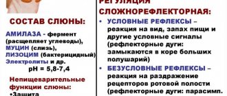
12.What is the role of saliva in digestion? What reflexes provide salivation and under what conditions?
1) saliva contains enzymes that break down starch, as well as substances that form a food bolus for swallowing; 2) unconditioned reflex salivation occurs when the receptors of the oral cavity are irritated; 3) conditioned reflex salivation occurs in response to irritation of the visual, olfactory, and auditory analyzers .
13. Find errors in the text. Indicate the numbers of sentences in which errors were made and explain them. 1. The stomach is the widest part of the digestive tract. 2. It is located above the diaphragm on the left side of the abdomen. 3. The gastric mucosa contains many glands. 4. Some of them secrete sulfuric acid, which activates the work of digestive enzymes. 5. These include pepsin, amylase and maltase.6.Food from the stomach enters the duodenum through the muscular sphincter.
Errors are contained in the sentences: 1) 2 - the stomach is located under the diaphragm; 2) 4 - the glands of the stomach secrete hydrochloric acid; 3) 5 - amylase and maltase are not enzymes of gastric juice. These are salivary enzymes.
14. How is neurohumoral regulation of gastric juice secretion carried out in the human body? Explain your answer.1.
nervous regulation is carried out by direct irritation of the receptors of the oral cavity and stomach (unconditioned reflex) 2. nervous regulation is carried out by irritation of the receptors of the visual, auditory, olfactory analyzers (conditioned reflex) 3. humoral regulation: products of the breakdown of organic substances formed in the stomach are absorbed into the blood and through blood affects the glands of the stomach (gastrin)
15. In a sheep, the length of the intestine is approximately 28 times greater than the length of the body, in a person - 6 times, and in a lion - only 3 times. What explains this difference?
1) Plant foods (fiber) are more difficult to digest in the digestive canal than animal foods. Consequently, the length of the intestine depends on the nature of the food consumed: the more plant food it contains, the longer the intestine. 2) The sheep eats exclusively plant food, and it has the longest intestine; The lion eats meat, and it is easily digested and assimilated. Man eats mixed food
16.What is the relationship between the structure and functions of the human small intestine?
1. the intestinal walls have glandular epithelium, the cells of which produce enzymes that promote digestion 2. intestinal villi greatly increase the area of the mucous epithelium, which ensures efficient absorption of nutrients
17.What is the significance of the large length of the small intestine for the digestion of food?
1) The long length of the small intestine allows the completion of the conversion of organic matter from food into soluble compounds 2) The long length of the small intestine facilitates the absorption of nutrients
18.What features of the inner surface of the small intestine ensure intensive absorption of nutrients in it?
1) Intensive absorption of nutrients is ensured by a large suction surface area, which is many times greater than the surface of the human body. 2) The absorption surface area increases due to the presence on the inside of the small intestine of a large number of villi and microvilli covered with epithelial tissue
19. In which part of the digestive tract do symbiotic microorganisms live, what function do they perform?
1 live in the large intestine 2 bacterial enzymes break down fiber, preventing the process of rotting 3 bacteria synthesize vitamins K and B12
20. A large number of bacteria live in the large intestine and constitute normal microflora, their
role.
1. break down fiber and also destroy unabsorbed protein digestion products2. synthesis of B vitamins, vitamin K.3. suppression of the proliferation of putrefactive and pathogenic bacteria
21.Why can treating a person with antibiotics lead to intestinal dysfunction? Give at least two reasons. 1
. antibiotics kill beneficial bacteria that live in the human intestines 2. the breakdown of fiber, water absorption and other processes are disrupted
22.What negative consequences for human health can the use of pesticides have to combat the Colorado potato beetle? Explain why.
1. poisons accumulate in potato tubers 2. eating such potatoes can cause poisoning and other diseases
23. What functions does the liver perform in the human and animal body?
1. produces bile, which is involved in the breakdown of fats; 2. it disinfects toxic substances that are formed in the body or come from food; 3. in the liver, glucose is converted and stored in the form of glycogen; 4. participates in the regulation of the metabolism of fats, proteins, vitamins, hormones and other biologically active substances;5. participates in the production of thrombin, fibrinogen5) is the site of destruction of red blood cells, blood depot functions
24.Functions of bile
1. emulsifies fats, which is necessary for their breakdown by pancreatic lipase 2. promotes the absorption of fat breakdown products in the small intestine 3. stimulates the secretory activity of the pancreas 4. neutralizes the acidic contents of the stomach entering the duodenum 5. enhances intestinal motility 6. promotes excretion a number of foreign and harmful substances to the body
25. What can cause vomiting?
1. entry of toxic substances into the blood 2. irritation of receptors in the mucous membrane of the digestive canal 3. conditioned reflex 4. diseases (hypertension, hepatitis)
26. In practice, it has been observed that when taking medications by mouth, their effect is weaker than when administered intramuscularly or into a vein. Give an explanation for this phenomenon.
1. when taking medications by mouth, some of them are destroyed in the digestive canal and in the liver 2. from the muscles or veins they are more likely to enter the internal environment
27.Can a person eat only fats, excluding protein foods from the diet?
1) It cannot. 2) The absence of proteins will lead to the fact that the following functions will not be performed in the cells: enzymatic, motor, protective, transport. 3) The synthesis of proteins from fats is impossible, because proteins have a more complex structure (their molecules include nitrogen and sulfur).
28.Why should the human diet include food of animal origin?
1) Plant proteins, unlike animals, do not contain all the amino acids necessary for the formation of proteins specific to the human body 2) Animal fats are closer in composition to human fats than plant fats
29. It is recommended to use “seaweed” - kelp - in the human diet. What is the significance of its use in normalizing body functions?
1) Laminaria is a brown algae that contains iodine. 2) Iodine is necessary for the normal functioning of the thyroid gland. Iodine is part of the hormone thyroxine, which in turn regulates metabolism and the functioning of the nervous system.
30.What measures are used to combat parasitic worms?
1. Compliance with personal hygiene rules2. Purification of drinking water3. Sanitary control in slaughterhouses and food preparation.
31.Where is the center of unconditioned reflex regulation of pancreatic juice located and what is the humoral regulation of this process? What is the role of this juice in digestion?
1) the center is located in the medulla oblongata
2) humoral regulation is due to substances entering the blood during the breakdown of food
3) pancreatic juice contains enzymes that break down proteins, lipids, and carbohydrates of food into their monomers, which can be absorbed by the body’s cells
32. People of many professions stand motionless on their feet throughout the working day, so they often develop an occupational disease - dilatation of the veins of the lower extremities. Explain why this is happening.
1) when standing for a long time, the outflow of blood from the veins is disrupted; 2) there is no contraction of the muscles of the lower extremities, which contribute to the contraction of the walls of the veins and the movement of blood upward
33.Where are the centers of food regulation of gastric juice located in the human body? How is unconditioned reflex and conditioned reflex regulation of digestive processes carried out?
1) the unconditioned reflex center is located in the medulla oblongata. Conditioned reflex - in the GM KBP2) the conditioned reflex center ensures the separation of gastric juice when food enters the oral cavity and stomach 3) Conditioned reflex release of gastric juice occurs at the sight, smell, thought of food
34. Find errors in the given text. Indicate the numbers of the sentences in which they are made, correct them. 1. In the oral cavity, food is crushed and moistened with saliva. 2. Saliva contains enzymes and substances that kill germs. 3. Saliva enzymes break down proteins into amino acids. 4. The esophagus produces enzymes that cause chemical changes in food. 5. The movement of food gruel occurs due to the contraction and relaxation of the muscles of the intestinal walls. 6. Absorption of the bulk of nutrients occurs in the cecum. Errors were made in the sentences:
3 - salivary enzymes do not act on proteins, but break down starch; 4 - enzymes are not produced in the esophagus, so their effect is excluded; 6 - absorption of the bulk of nutrients occurs in the small intestine.
35.What is the role of saliva in digestion? What reflexes provide salivation and under what conditions?1) saliva contains
enzymes that break down starch, as well as substances that form a food bolus for swallowing; 2) unconditioned reflex salivation occurs when the receptors of the oral cavity are irritated; 3) conditioned reflex salivation occurs in response to irritation of the visual, olfactory, and auditory analyzers.
36. Indicate the functions of the large intestine of mammals and humans.1
. Formation of burl masses
2. Suction of most of the water3. Normalization of digestion and absorption due to bacterial flora 4. Carrying out the act of defecation.5. bacteria break down fiber and also destroy unabsorbed protein digestion products, ensure the synthesis of B vitamins, vitamin K, suppress the proliferation of putrefactive and pathogenic bacteria
37. Biological scientists measured the length of the digestive system of a predatory and herbivorous animal of equal size. Which of these animals had a longer digestive tract and why?
A plant cell, in addition to the plasma membrane, also has an outer cellulose membrane that protects it from the influence of environmental factors. This membrane is more difficult (and takes longer) to destroy with the help of digestive enzymes than the plasma membrane of an animal cell. Therefore, animal food is digested faster than plant food.
38)
Functions of bile
1. emulsifies fats (turns them into droplets), which is necessary for their breakdown by pancreatic lipase (i.e. activates lipase)
2. promotes the absorption of fat breakdown products in the small intestine
3. stimulates the secretory activity of the pancreas
4. neutralizes the acidic contents of the stomach entering the duodenum
5. enhances intestinal motility
6. helps eliminate a number of substances that are foreign and harmful to the body
39) Why should food be chewed thoroughly before swallowing?
- Chewing food is a mechanical process that increases the surface area of contact with saliva.
- Salivary enzymes help break down complex carbohydrates into simple ones, and lysozyme disinfects food.
Pathological anatomy
Dystrophic changes in the salivary glands are often combined with a violation of their functions. Protein dystrophies (see Protein dystrophy) are characterized by cloudy swelling of glandular cells (granular dystrophy) and hyalinosis of interstitial tissue (see Hyalinosis). Granular degeneration of glandular cells is observed with sialadenitis (see), cachexia (see), as well as with poisoning with salts of heavy metals (mercury, lead, etc.), released with saliva and damaging glandular cells. Hyalinosis of the interstitial tissue leads to thickening of the interlobular septa; hyaline can be found in the walls of small vessels and in the basement membranes of the terminal (secretory) sections of the veins. With general amyloidosis (see), amyloid is occasionally deposited in the walls of blood vessels and basement membranes. Fatty degeneration of glandular cells (see Fatty degeneration) is observed in infectious diseases (diphtheria, tuberculosis) and chronic cardiovascular diseases. Lipomatosis S. is expressed in the growth between their lobules of adipose tissue (see Lipomatosis). Excessive development of adipose tissue in the thickness of the stomach. occurs in general obesity (see) and senile atrophy of the stomach.
Carbohydrate dystrophies (see) are observed in diabetes mellitus and are characterized by the appearance of inclusions of glycogen (see) in the cytoplasm of glandular cells.
Necrosis S. g. is rare and occurs. arr. with purulent denitis.
Disorders of blood and lymph circulation in the stomach. have no independent significance in the overall pathological picture. They are only local manifestations of general circulatory disorders, occurring in the form of venous hyperemia (see). Arterial hyperemia is observed, as a rule, during local inflammatory processes in the stomach. and in case of disruption of innervation. Hemorrhages in the S. g. are noted in blood diseases, injuries and certain inf. diseases (eg, typhoid and typhus).
An acute nonspecific inflammatory process in the gland—adenitis—occurs as a result of the introduction of pathogens of a viral or bacterial infection that penetrate the gland through the excretory duct, as well as by hematogenous or lymphogenous routes. There is a diffuse form of inflammation of the stomach. and limited (in the form of an abscess). Chronic nonspecific sleeping adenitis can occur primarily and less frequently, as an outcome of acute inflammation of the stomach. With chronic inflammation of the stomach decrease in size and acquire a dense consistency. Microscopically, infiltration of the stroma and parenchyma of the glands with lymphoid elements, atrophy of the terminal (secretory) sections and pronounced stromal sclerosis are noted.
Defeat S. zh. in tuberculosis it manifests itself in the form of a unilateral lesion and occurs secondarily as a result of lympho- and hematogenous dissemination. Development of tuberculosis S. begins with damage to regional lymph nodes of the gland, subsequently its parenchyma and stroma are involved in the process. In S. zh. detect miliary tubercles or small nodules undergoing caseous necrosis. Sometimes there is melting of caseous masses with the formation of a tuberculous abscess, which can cause fistulous tracts. With histol. The study reveals tuberculous tubercles of a normal structure (see Tuberculosis), and when the tissue melts, nonspecific inflammatory changes occur.
Defeat S. zh. It is rare in syphilis. Inflammatory changes are detected only in the secondary and tertiary periods of the disease and can be diffuse or limited in nature. With diffuse inflammatory infiltration (most often in the secondary period) S. It is affected throughout, and abscess formation and necrosis of individual areas of the gland often develop. Limited forms of damage are characterized by sclerosis and atrophy of the stomach. Histologically in the S. g. detect changes corresponding to the picture of specific inflammation in syphilis (see).
Actinomycosis of the salivary glands can develop primarily, as a result of the penetration of actinomycetes into the gland along the ducts, or secondarily, when the inflammatory process spreads from the surrounding salivary glands. soft tissues. Primary actinomycosis (see) proceeds slowly, with periodic exacerbations. In S. zh. a dense, limited infiltrate is revealed, which subsequently spreads to the entire gland with the formation of multiple abscesses. Secondary actinomycosis has less clear symptoms due to the development of a diffuse inflammatory process in the surrounding tissues. Specific inflammatory changes in actinomycosis of S. characterized by the development of granulation tissue with small pustules and pronounced signs of scarring. Druses of actinomycetes are often found in the inflammatory infiltrate, which, when localized in the excretory ducts, can be the basis for the formation of salivary stones.
Of the parasitic diseases of S. zh. Cases of echinococcosis are extremely rare. At the same time, S. are affected secondarily, and macro- and microscopic changes in them are similar to those with damage to other organs (see Echinococcosis).
Hypertrophy of the stomach. is a response to patol. processes occurring in the body. Increase in s. observed in endocrine diseases (eg, diffuse toxic goiter, hypothyroidism), cirrhosis of the liver and usually occurs as a result of reactive proliferation of interstitial tissue, which leads to interstitial sialadenitis. Hypertrophy of interstitial tissue is also observed in Mikulicz syndrome (see Mikulicz syndrome). In physiol. conditions of hypertrophy of the stomach. observed during pregnancy and the postpartum period. Sometimes, after removal of one of the paired glands, vicarious hypertrophy develops on the opposite side.
Atrophy of the s. characterized by a decrease in their size. Atrophic changes are observed when there is a violation of the innervation of the gland, age-related involution, as well as when the outflow of gland secretions is difficult, followed by atrophy of the parenchyma. Histologically, there is a proliferation of connective tissue with thickening of the interlobular septa, a decrease in the size of glandulocytes, and emphasized lobulation of the sternum.
Postmortem changes in S. zh. occur early (after 3-4 hours), which is associated with the self-digesting effect of salivary enzymes. Macroscopically, the glands acquire a reddish tint and soften. With pathohistol. The study reveals destructive changes in glandular cells, while interstitial tissue retains its structure much longer.
Examination methods include, in addition to general methods (questioning, examination, palpation, etc.), such special methods as probing the ducts, sialometry (see Salivation), cytol. examination of secretions, ultrasonic dowsing (see Ultrasound diagnostics), thermovisiography (see Thermography), scanning (see), sialography (see), pantomography (see), pneumosubmandibulography (see), computer tomography (see).
Development[edit]
| This section needs expansion . |
Aging[edit]
With aging of the salivary glands, some structural changes are observed, such as: [21] [22]
- Reduction of acinar tissue volume
- Increased fibrous tissue
- Increase in adipose tissue
- Hyperplasia and dilatation of ducts [21]
In addition, changes in saliva content are also observed:
- Decrease in the concentration of secretory IgA [21]
- Reducing the amount of mucin
However, there is no overall change in the amount of saliva produced.
Clinical significance[edit]
Microphotograph of chronic inflammation of sialadenitis of the salivary gland).
Main article: Salivary gland disease
Sialolithiasis (salivary calculus or stone) can cause blockage of a duct, most commonly the submandibular duct, causing pain and swelling of the gland. [32]
Salivary gland dysfunction refers to either xerostomia (a symptom of dry mouth) or salivary gland hypofunction (decreased saliva production); it is associated with a significant deterioration in quality of life. [33] Following radiation therapy to the head and neck region, salivary gland dysfunction is a predictable side effect. [33] Saliva production can be stimulated pharmacologically using salivary agents such as pilocarpine and cevimeline. [34] It can also be suppressed by so-called antisialogic drugs, such as tricyclic antidepressants, SSRIs, antihypertensives, and polypharmacy. [35] A Cochrane review found that there is no convincing evidence that topical treatments are effective in relieving symptoms of dry mouth. [36]
Cancer treatments, including chemotherapy and radiation therapy, can interfere with the flow of saliva. [36] [33] Radiation therapy may cause persistent hyposalivation due to damage to the oral mucosa containing the salivary glands, leading to xerostomia, whereas chemotherapy may cause only temporary impairment of salivary function. [36] [33] In addition, surgical removal due to benign or malignant lesions can also impair function. [37]
Graft-versus-host disease after allogeneic bone marrow transplantation may present with dry mouth and many small mucoceles. [38] Tumors of the salivary glands may occur, including mucoepidermoid carcinoma, a malignancy. [39]
Clinical trials/research[edit]
A sialogram is a radiopaque study of the salivary duct that can be used to examine its function and to diagnose Sjögren's syndrome. [40]