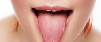
Anatomy of the tongue
The tongue is divided into: body, apex and root. The tongue consists of muscles and is covered with epithelium. Also on the back of the tongue there are many special papillae with which we feel taste. The largest grooved papillae are located closer to the root of the tongue. In the center of the tongue there are filiform papillae, on the sides there are leaf-shaped papillae. And in front of the tongue there are mushroom-shaped papillae.
What it is
The natural microflora of the oral cavity involves the presence of a mass of bacteria, viruses and fungi, which, with a good level of body resistance, are in a “dormant” state or controlled by beneficial bacteria. As soon as any organs malfunction and the immune system drops sharply, all pathogenic microflora are activated and begin to multiply intensively.
Fungi of the genus Candida are no exception. Exceeding their number above the natural norm (it does not matter whether they came from the outside or their proliferation was a consequence of antibiotic therapy) is the main cause of the development of candidal glossitis. Thus, the inflammatory process with the formation of a white cheesy coating on the back of the tongue is the first manifestation of this disease.
Those most at risk are newborns and infants with undeveloped immunity, elderly people with a large number of chronic diseases, women of childbearing age and men who smoke frequently. In addition, in people with diabetes, oncology and HIV, candidal glossitis appears very often.
The infection enters the oral cavity through direct contact with infected people (kissing or sharing utensils), or when a child is breastfed by an infected mother. The possibility of fungus entering the body through water, dirty objects and food, blood transfusions, using non-sterile reusable medical instruments, as well as through the unwashed hands of medical personnel (for example, in maternity hospitals) cannot be ruled out.
Glossitis: the cause of the disease
Tongue diseases can be caused by infections caused by:
- Bacteria (streptococci, staphylococci, fusobacteria, etc.)
- Mushrooms
- Viruses
The tongue is often affected by common diseases:
- immune system diseases
- blood diseases
- diseases of the digestive system
- with a lack of vitamins, microelements (for example, iron and B12)
Also, the cause of glossitis can be irrational use of antibiotics, which disrupts the microflora of the oral cavity, or bad habits (smoking, alcohol).
Glossitis can occur due to tongue injuries:
- sharp edges of carious or damaged teeth, fillings
- old dentures, crowns
- when biting with teeth
- fish bones and other items
- gunshot and other types of injuries
- chemical irritants (for example, vinegar)
- hot food
- electric shock
Classification of glossitis according to the form of the lesion
- catarrhal form
- desquamative form
- purulent form
- neurogenic diseases of the tongue (impaired sensitivity of the tongue)
Catarrhal form of glossitis
With this form, only the upper layers of the tongue are affected. Areas of redness and swelling appear on the tongue. The tongue is coated, a burning sensation appears, the amount of saliva increases, and the mobility of the tongue is sometimes limited
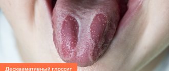
Desquamative form of glossitis
With this form, only the upper layers of the tongue are affected. Areas of redness and swelling appear on the tongue. The tongue is coated, a burning sensation appears, the amount of saliva increases, and the mobility of the tongue is sometimes limited
Purulent form of glossitis
Its cause is infection, most often through a damaged mucous tongue.
It is characterized by a violation of the general condition, weakness, temperature 38-39°C, and enlarged lymph nodes. The tongue increases in volume and is covered with a gray coating, the saliva is viscous. Bad breath appears, it is difficult to swallow food, and even breathing may be difficult. If you notice the above symptoms, you should urgently seek medical help!
Separately, there are diseases in which only the tongue is affected without the rest of the oral mucosa:
- Desquamative glossitis
- Black hairy tongue
- Folded tongue
- Diamond-shaped glossitis
- Neurogenic diseases of the tongue
Hairy tongue
With this disease, filiform papillae grow and become black. Coloring occurs due to food and microflora. The disease is rare.
Desquamative glossitis
With this disease, the process of keratinization and changes in the papillae of the tongue are disrupted. More common in children. Part of the tongue is covered with red spots that can migrate to other areas. The disease is accompanied by dryness and impaired taste.
Diamond-shaped glossitis
A chronic disease of unknown cause in which a red or bluish diamond-shaped formation is located in the middle of the tongue in front of the circumvallate papillae. Usually there are no complaints, rarely dryness, pain, burning.
Folded tongue
A congenital disorder of the structure of the tongue, with it there are folds on the tongue, often symmetrical. Sometimes the disease is accompanied by an enlarged tongue. No subjective unpleasant sensations arise; only with insufficient oral hygiene, microorganisms can develop in the folds of the tongue, causing diseases (for example, candidiasis).
Neurogenic diseases of the tongue
Violation of the motor and trophic (nutritional) function of the tongue. They are expressed in impaired sensitivity, mobility, pain, as well as in impaired desquamation (detachment).
Membranoprotective therapy for the allergic form of desquamative glossitis
K. G. Karakov Doctor of Medical Sciences, Professor, Head of the Department of Therapeutic Dentistry of St. State Medical Academy
T. N. Vlasova Associate Professor, Department of Therapeutic Dentistry, St. State Medical Academy
A. V. Oganyan Candidate of Medical Sciences, Assistant of the Department of Therapeutic Dentistry of St. State Medical Academy
O. V. Polyakova Clinical Resident, Department of Therapeutic Dentistry, St. State Medical Academy
Desquamative glossitis is a disease of the mucous membrane of the tongue, leading to the formation of areas of desquamation (flaking, peeling) of the epithelium on its surface.
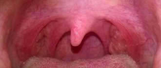
Desquamation areas have different shapes and are located both on the back of the tongue and on its lateral surfaces (Fig. 1-4).
Rice. 1
Rice. 2
Rice. 3
Rice. 4
The disease occurs quite often, its occurrence does not fundamentally depend on the gender and age of a person.
Currently, there are no clearly defined causes for the development of desquamative glossitis. It is known that the development of the disease is based on a malnutrition of the tissues of the mucous membrane of the tongue, as a result of which they desquamate. Difficulties lie in determining the reasons for the violation of trophic tissue of the tongue.
It has been determined with a high degree of confidence that the causative factors for the appearance of desquamative glossitis are diseases of the gastrointestinal tract, pathology of the nervous, hematopoietic, cardiovascular and endocrine systems.
The trigger for the development of desquamative glossitis may be an allergic predisposition, hypovitaminosis, or connective tissue diseases. Desquamative glossitis often accompanies candidiasis (Fig. 5).
Rice. 5
Clinically, superficial, hyperplastic and lichenoid forms of desquamative glossitis are distinguished.
The superficial form is characterized by the appearance of well-demarcated red stripes and spots on the dorsal surface and edges of the tongue. The spots have the appearance of raw meat and are surrounded by intact mucous membrane of normal color. With diffuse desquamation of the epithelium of the tongue, its surface becomes smooth and shiny (“polished tongue”).
This area stands out against the surrounding slightly raised, non-desquamated zone of opacified epithelium. The affected area quickly spreads along the periphery, but retains smooth, rounded boundaries. The intensity of desquamation gradually decreases. Subjectively, itching and burning are noted. Patients complain of an unusual appearance of the tongue.
The hyperplastic form of desquamative glossitis is characterized by focal thickening of the filiform papillae of the tongue, the presence of white, yellow, gray or dark plaque in the affected area, accompanied by a sensation of a foreign body.
The lichenoid form of desquamative glossitis is characterized by the appearance of areas of desquamation of the epithelium of the tongue of various shapes and sizes due to the redistribution of filiform papillae and their grouping surrounded by exposed areas. In the desquamation zone, the fungiform papillae are enlarged. The pattern of changes in the surface of the tongue is not permanent; foci of desquamation can move along the surface of the tongue.
Shumsky A.V., Burda A.G. identify the following forms of desquamative glossitis:
- dysbiotic (microbial) is characterized by the presence in fingerprint smears of pathogenic (St. aureus, β-hemolytic Str. group A) and conditionally pathogenic (staphylococci, clostridia, corynebacteria, etc.) flora;
- candidiasis is characterized by a predominance of blast forms of fungi of the genus Candida with oval and round blastospores, as well as a large number of young and mature forms of pseudomycelium;
- neurogenic (increased norepinephrine concentration to 2.37 ± 0.64 when the norm is 0.35 ± 0.12 ng/ml, ρ ‹ 0.05);
- allergic (eosinophilia, histamine content 0.94 ± 0.08 ng/ml, normal 0.40 ± 0.08 ng/ml).
We are most interested in the allergic form of desquamative glossitis.
The purpose of our study was to study the membrane-protective properties of modern immunocorrective and antihistamine drugs in the treatment of the allergic form of desquamative glossitis.
We examined and treated 7 people with an allergic form of desquamative glossitis aged from 25 to 60 years. All examined were divided into 2 groups: main and control. Main group: 4 people, two men and two women. Control group: 3 people, two men and one woman. The duration of the disease varied from several weeks to 2-3 years.
The clinical examination included the identification of complaints, study of life history and disease, visual examination of the oral cavity and tongue, palpation of the tongue and regional lymph nodes, microbiological examination to assess the contamination of the oral mucosa and dorsum of the tongue with Candida fungi and other microorganisms.
The examination was carried out by dentists together with allergists and gastroenterologists. All examined patients had a history of allergic status and pathology of the gastrointestinal tract.
When examining the oral cavity of patients with an allergic form of glossitis, the mucous membrane was pale and moderately moist. There were tooth marks on the lateral surfaces of the tongue, which indicated interstitial edema. Microscopy of the smear-imprint was practically no different from the microscopic picture in healthy individuals.
Treatment regimens
Patients of the main group received standard therapy:
Local:
- sanitation of the oral cavity;
- antiseptic rinses with chlorhexidine solution;
- applications to the lesions of keratoplasty preparations (solcoseryl jelly, rosehip oil, vitamin A).
General treatment included elimination of the allergenic factor (if possible), vitamin therapy, the use of an antihistamine (Diazolin), 1 tablet 2 times a day for two weeks, an immunocorrective drug (Imudon), 2 tablets 6 times a day for within two weeks.
Patients in the control group received standard therapy using the latest generation of desensitizing drugs (Tinset, 1 tablet 2 times a day for two weeks) and immunocorrective drugs (Likopid, 2 tablets 2 times a day sublingually for two weeks).
During the study, we received a stable remission - a small number of complaints, a decrease in lesions in all those examined. But in the control group, the remission phase began after the 6th visit, and in the main group - by the end of the second week.
The study of the desquamative form of glossitis against the background of an allergic status and the use of modern drugs allowed us to achieve more stable remission in the control group compared to the main group, and therefore we recommend the use of this regimen in practical healthcare.
- Banchenko G.V., Maksimovsky Yu.M., Grinin V.M. Language is the “mirror” of the body: Clinical guide for doctors. - M., 2000. - 407 p.
- Banchenko G.V. Combined lesions of the oral mucosa and internal organs. - M.: Medicine, 1979.
- Burda A.G. Pathogenetic rationale for complex treatment of desquamative glossitis: Abstract of thesis. dis. Ph.D. honey. Sci. - Samara, 2006.
- Danilevsky N. F. et al. Oral diseases. - M., 2001. - 271 p.
- Rybakov A. I., Banchenko G. V. Diseases of the oral mucosa. - M.: Medicine, 1971.
- Yamashev I. G., Solovyov M. M. Linguology as a scientific direction // Kazan. Vestn. dentist - 1996, No. 2. - P. 106-108.
Diagnosis of glossitis
If symptoms of glossitis appear, you should consult a dentist.
The doctor will collect the entire history of the disease, complaints, duration of the disease, the success of previous treatment (if any). The doctor examines not only the tongue, but also the entire oral cavity, including teeth, crowns, and dentures.
In some cases, additional research may be needed:
- Ultrasound examination of the salivary glands
- Smear to identify the pathogen and select appropriate treatment
- Bacteriological culture of saliva
These studies are needed to find the cause of the disease and prevent complications.
How to treat glossitis?
Treatment of glossitis is always comprehensive and carried out by a dentist. Also, if necessary, the help of a therapist, allergist, gastroenterologist, ENT doctor, endocrinologist is required.
Treatment depends on the course and form of the disease and includes the following measures:
- Elimination of pockets of infection in the mouth (treatment of caries, its complications, replacement of old fillings, crowns)
- Professional oral hygiene – removing plaque and tartar to reduce irritation and reduce the number of microorganisms.
- Teaching the patient how to properly brush teeth at home, selecting appropriate products (paste, brush, brushes)
- Gentle diet (spicy, salty, hot, irritating foods must be excluded)
- If there is a general disease, it must be cured
Medicines for the treatment of glossitis
- Antibiotics
- Antifungal medications (to treat fungal infections)
- Antiviral medicines (to treat a viral infection)
- Immunomodulators (to enhance immune system function)
- Antihistamines (to reduce tongue swelling)
- Vitamins
- Applications of healing agents to lesions (Solcoseryl, rosehip oil, vitamin A)
All of these forms of drugs are selected individually, taking into account sensitivity to the identified pathogens. The duration of treatment is determined by the doctor in accordance with the form and severity of the disease.
Prevention of glossitis
- Timely diagnosis and treatment of common diseases. To do this, it is recommended to undergo regular medical examinations.
- Regular and proper brushing of teeth at home twice a day.
- Visit the dentist once every six months for examination and professional hygiene (if necessary).
- Healthy lifestyle. It is important to eat food with sufficient protein, fat, and carbohydrates. It is better to minimize irritating foods (smoked, salted). It is also necessary to normalize your sleep patterns.
- Rejection of bad habits. Tars in tobacco smoke irritate the mucous membrane of the tongue and also impair blood supply.
How and with what to treat glossitis of the tongue in adults and children
Treatment of tongue glossitis requires elimination of the diseases that led to its appearance. This refers to anemia, gastrointestinal diseases, syphilis, etc. To minimize pain, you need to rinse your mouth with Furacilin, a solution of Chlorhexidine (0.05%) or potassium permanganate, and avoid eating rough food. In case of severe pain, local applications with anesthetics can be made:
- Trimecaine solution (2%);
- Lidocaine solution;
- Pyromecaine solution.
If the tongue is dry, it should be lubricated with a mixture of glycerin and Anestezin.
For a speedy recovery, the skin of the tongue must be cleaned of plaque using a cotton swab soaked in proteolytic enzymes (Chymotrypsin, Trypsin). If there are painful ulcers, these drugs for the treatment of tongue glossitis can be replaced with applications with Iruksol. After the hygiene procedure, it is important to treat the cleaned lesions with antiseptics. This helps prevent secondary infection and also prevents the development of serious complications.
To speed up regeneration processes, gel and jelly-like medicines with solcoseryl, as well as preparations containing vitamin A, can be used. The mixture formed by rosehip oil and vitamin A heals the affected skin well and relieves pain.
If excessive thickening of the stratum corneum of the epidermis (hyperkeratosis) is observed, surgical intervention is ordered. Antifungal, antibacterial and anti-inflammatory drugs are selected taking into account the severity of the disease and the dominant symptoms.
All patients with glossitis can take immunomodulators. Hormones are used extremely rarely - only for difficulty breathing. To avoid atrophy of taste organ cells, Hydrocortisone and Prednisolone ointments are prescribed in short courses.
How to treat tongue glossitis at home
Treatment of tongue glossitis at home can be carried out using traditional medicine recipes:
- decoction of chamomile flowers. 1 tbsp. pour a glass of boiling water over the flowers. Infuse, strain. Rinse your mouth after every meal;
- bedstraw decoction. 1 tbsp. pour a glass of boiling water. Leave for 30 minutes. Take 50 ml orally after meals and rinse the mouth 3-4 times a day. Similarly, you can use decoctions of sage and basil;
- soda solution. Add 2-3 drops of iodine and a teaspoon of baking soda to a glass of warm water. Rinse your mouth 3 times a day;
- natural honey Slowly dissolve a spoonful of honey;
- propolis. Lubricate the areas of inflammation 5 times a day. Instead of propolis, you can use raw carrot/potato juice or rosehip oil;
- tea tree oil. Dilute in equal proportions with olive, sunflower or sea buckthorn oil. Treat glossitis lesions 5 times a day or keep it in your mouth for several minutes.
Answers to popular questions about glossitis
Do I need to clean my tongue?
Yes, definitely.
A lot of microorganisms accumulate on the tongue, which leads to bad breath. In case of acute glossitis, the primary task is to relieve inflammation; you can use oral baths or rinses with antiseptic solutions (chlorhexidine, miramistin), a decoction of chamomile has a positive effect (has a calming effect on inflamed tissues of the mucous membrane of the tongue and the oral cavity as a whole). Also, do not forget about careful cleaning of the surface of the tongue.
There are special scrapers (semi-oval) for cleaning the tongue. They are designed to gently cleanse the tongue. Do not clean your tongue with brushes or spoons, as this can cause injury.
Which doctor should I contact to treat glossitis?
If tongue pathology is detected, you can consult a dentist. And, if necessary, the dentist will refer you for consultation to other specialists if this is due to the presence of a general disease.
What diagnostics are carried out before treating glossitis?
The specialist to whom the patient contacted conducts an examination of the oral cavity, collects a medical history, data on the general condition of the body (the presence of chronic organ diseases, etc.), prescribes tests, as well as laboratory diagnostics: a smear or culture of the oral cavity and tongue to determine pathogenic microflora.
Are glossitis inherited?
Yes, some forms can be inherited, for example, a folded tongue.
Is it possible to completely cure glossitis?
Some forms can be successfully treated, but often glossitis is a chronic and congenital disease. But with proper therapy, you can achieve minimal manifestations of the disease.
What is desquamative glossitis?
The surface of the tongue is covered by a mucous membrane lined with stratified epithelium. Normally, it is pink and uniform in color. With desquamative glossitis, the epithelium begins to exfoliate and slough off, forming red spots of varying sizes with a light border. Inflamed areas can cause pain and a burning sensation, but often the disease is not accompanied by discomfort, so the patient may not notice it at all.
Foci of inflammation periodically change their location: the epithelial layer is restored in one area, then destroyed in another. Thus, the spots constantly “migrate”, and the language becomes like a geographical map. This is where desquamative glossitis gets its informal name.